
What Is An Ultrasound of Carotid Artery?
Ultrasound is an imaging technique used to examine internal organs. Carotid ultrasound is a non invasive procedure for assessing the carotid arteries in the neck. Ultrasound uses high frequency sound waves and creates an echo once they hit the target object. Based on the echo a trained technician performing the test is able to determine the shape and size of the organ, detect any unusual changes, and spot masses of cells that could be tumors. The carotid arteries are positioned one on each side of the neck. Their primary aim is to transport blood from the heart into the brain. A special type of ultrasound called the Doppler Effect is used to assess the condition of arteries, veins, and blood vessels. Using the Doppler Effect medical care professionals inject a harmless dye intravenously into the patient and follow it as it makes its way from organ to organ via blood stream. Any blockages in the organs or the circulation system show on the ultrasound. In addition, carotid ultrasound is used to diagnose disorders, propose treatment, and assess the outcome of therapy. Compared to X-rays which emit radioactive waves, ultrasound is safe to use as it does not produce any adverse health effects.The Purpose of Carotid Ultrasound
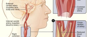
Health care providers will send an individual for a carotid ultrasound if they suspect one of the arteries may be narrowing, which could result in a stroke. Narrowing of any blood vessels is caused by deposits of fat, cholesterol, and calcium. Using ultrasound, the doctors are able to diagnose problems early and propose courses of treatment to stimulate the blood flow. Patients who suffer from underlying illnesses which increase the possibility of a stroke are likely to be referred for an ultrasound. Individuals who have been battling high cholesterol will be advised to have periodic carotid ultrasound. Diabetes and high blood pressure also increase the risk of stroke and it is recommended that the condition of the blood vessels be monitored regularly. Finally, family history of stroke or heart problems predisposed the individual to be affected by similar types of disorders so regular exams are commended. In order to get the most valid results, carotid ultrasound is often combined with other types of tests. For instance, ankle-brachial index test is performed to compare the blood pressure at a person’s ankle and at their arm to screen for any blockages of blood flow to the legs. Abdominal ultrasound is also often administered to evaluate the abdominal aorta and make sure there are no aneurysms, and if there are to provide treatment immediately. Other than to assess the risk of a stroke, carotid ultrasound is also done to evaluate the structure of the arteries which might be deformed and in turn disrupt blood flow. Blood clots are also visible through ultrasound and detecting them is vital for saving lives. Carotid ultrasound is also used to assess the effects of surgery. In patients suffering from circulation problems mesh tubes are inserted to support weakened arteries and increase the blood flow. In other instances patients undergo surgery to remove the build up in arteries. The outcome of the procedure is easily determined via ultrasound.How Is The Procedure Done?
Preparation for a carotid ultrasound is fairly straightforward. Patients are advised to call the day before and confirm their appointments. They should wear comfortable clothing and no jewellery. Food and drink should be consumed as normal, and any prescribed medications need to be taken regularly unless otherwise specified. The ultrasound is performed by a trained technician or a radiologist. The equipment consists of a transducer, video screen, computer, and electronics. The transducer is rubbed against the neck to emit the sound waves and produce the images on the screen. The results are provided in real time and are available immediately. The technician will probably discuss them with a patient and send a formal report to the referring physician within a couple of days. The test should not last more than half an hour. In addition, the patient will be asked to lie on a bed for the duration of the procedure. The technician will apply clear gel on the neck to facilitate the process. Ultrasound tests should not produce any discomfort and therefore should be painless. If the patient is feeling uncomfortable the technician should be made aware.After The Ultrasound
Based on the results from a carotid ultrasound medical care professionals are usually able to set a diagnosis and prescribe treatment. If the results are not clear more tests will be ordered. Individuals whose results show that they are at an increased risk for a stroke will need to take certain steps. It is very important that the diet is light and healthy and that the individual is engaging in regular physical activity. Some patients will be prescribed medications to prevent blood clots and high cholesterol. In more serious cases surgery will be required to remove the build up from the arteries or to support the arteries with a mesh tube.

-Disease-Cause-A-Stroke_f_280x120.jpg)








Your thoughts on this
Loading...