The vein is a part of the circulatory system in charge of carrying deoxygenated blood towards the heart. There are only two exceptions. Namely, the umbilical veins (present only in fetuses) and the pulmonary vein actually carry the blood rich in oxygen. Now, the veins in legs collect the blood from these extremities and transfer it to the heart. Their structure and function can be easily examined by ultrasound. Ultrasound of veins in the legs is an integral part of diagnosing certain conditions such as deep vein thrombosis. This is a painless procedure, does not last long and provides with sufficient data for confirmation of certain abnormalities.
What is Venous Ultrasound?
Ultrasound imaging (also known as sonography) is a specific technique that visualizes the tissues exposed to high-frequency sound waves. The physics of sound usually places limits on the test so not all the tissues can be perfectly visualized with ultrasound. The quality of the images generally depends on many factors. For instance, images are poor in obese individuals in whom sound waves simply cannot penetrate deeply. Also, the exam is not efficient enough (or completely inefficient) when gas is present between the probe and the target organ. Ultrasound also does not visualize bone tissue.
This noninvasive medical test is highly suitable for visual perception of veins throughout the body. Apart from standard ultrasound of the veins in the lower extremities, this technique may be additionally used in the form of Doppler ultrasound, the exam that successfully evaluates blood flow through blood vessels.
Patients who are suspected of suffering from blood clots in deep veins of the legs are supposed to undergo venous ultrasound exam. The exam will either confirm the presence of blood clots or rule them out. Blood clots may easily dislodge and move towards the heart and the lungs triggering pulmonary embolism. This is a severe and potentially life-threatening complication of deep vein thrombosis which requires immediate medical help.
Furthermore, venous ultrasound study may be indicated in evaluation of prolonged leg swelling, varicose veins and is of great assistance when there is a need of a needle or catheter insertion. Ultrasound also assists in mapping the veins in the legs whose parts might be removed and then used as a bypass of blocked/narrowed blood vessels in other parts of the body (predominantly the heart.). And finally, the exam evaluates the blood vessel used for dialysis when there are problems with its functioning (e.g. narrowing).
Doppler ultrasound identifies blood clots, confirms or rules out blockage/narrowing and is of great help in the diagnosis of vascular tumors and congenital vascular malformations.
How is Procedure Performed?
There is no specific preparation for the exam. The clothes should be removed because the probe is in direct contact with the skin.
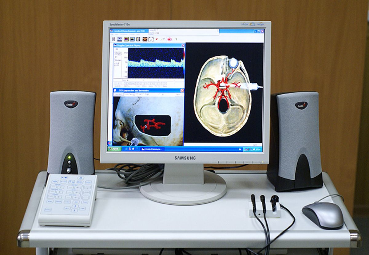
The very device comprises a console (a computer and electronics), a video display screen and a probe. The probe, also known as a transducer, is a tiny device which is held by the doctor and placed directly onto the patient's skin. There are several types of probes depending on the type of ultrasound exam. Some may be easily inserted in body cavities while others are designed to send and receive ultrasound waves through the skin. The principle is basically the same. The ultrasound waves are released from the probe, they penetrate certain tissues, are partially absorbed and partially refracted and again received with the probe. The images are directly displayed on the video screen. Doppler ultrasound evaluates the blood flow through the inspected veins. This approach allows doctors to easily notice blockage of any kind.
During the exam patients lie on their abdomen on an examination table. The table can be tilted or moved in other way to be adjusted according to doctor's needs.
Prior to examination, the area which is going to be in direct contact with the probe is covered in clear, water-based gel. The gel makes the probe stick to the skin and prevents any air pockets to disrupt the images. The radiologist performing the exam circles with the probe and keeps the probe firm to the skin right above the location of interest. The same probe is used for Doppler sonography. After the exam, the gel is removed from the skin and patients should wait for a while to receive doctor's report. The report is also sent to the physician who has referred the patient for the exam. The examination does not last long, between 30 and 45 minutes on average.
Since ultrasound is not an invasive technique it is clear that it does not hurt. The method is completely painless and causes no discomfort. Even when the probe is pressed firmly against the skin there is no pain. Pain might be reported and increase only if it has existed before the exam.
All in all, ultrasound is a rather quick, noninvasive diagnostic tool used in evaluation of many soft tissues and solid organs in the body. One of its many used is evaluation of blood vessels in the legs, veins in particular. With a standard ultrasound of the legs as well as Doppler ultrasound conditions affecting both superficial and deep veins in legs can be quickly diagnosed and treated almost instantly.


Your thoughts on this
Loading...