Granulomatosis with Polyangiitis (Wegener’s Granulomatosis) Overview
Granulomatosis with polyangiitis (formerly known as Wegener’s granulomatosis) (GwP) is an inflammation of the blood vessels of the lungs, kidneys, and other organs in the body. The disease is serious (and in some cases even systemic), and there are no recognized causes of the disease itself. The condition is life-threatening because it damages all affected organs in the body.
Estimated survival of GwP patients is about 87%, five years after the established diagnosis. Mortality may be caused by the disease or by the medications used for the treatment of the disease.

Patients usually consult the ophthalmologist because of the eye problems they keep experiencing, and this doctor is the first one that should suspect the GwP. An ophthalmologist can establish the diagnosis after he/she has seen the necrotizing scleritis and done some lab testing to confirm it.
Laboratory testing might include blood tests, urine analysis, chest X-ray, and some special testing, such as cANCA, computerized tomography, renal function tests, nasal and orbital biopsy, and c-reactive protein tests. Sometimes, necrotizing scleritis might be caused by some infections, and conjunctival scraping may be done to rule out this possibility.
In some cases, doctors might suspect that the patient is suffering from tuberculosis or rheumatoid arthritis. In those cases, the presence of progressing scleritis, orbital disease, and/or corneal problems, accompanied by some multisystem health problems will indicate Granulomatosis with polyangiitis disease.
The therapy requires a multisystem approach to every patient suffering from this disease. Corticosteroids are the first choice to treat GwP, but they don’t work alone, and they are not sufficient to stop the disease. Usually, doctors recommend a combination of corticosteroids and immunosuppressives to control the progression of the disease. The most commonly used medications are intravenous methyl-prednisolone and cyclophosphamide, and after that, oral use of prednisolone and cyclophosphamide.
Potential Health Problems
Granulomatosis with polyangiitis may cause problems with many organs in the human organism. The eyes, complete respiratory tract, and the kidneys are most commonly affected organs, but there is also a case of isolated lung or airway complications (in the limited form of the GwP). If the patient is suffering from the complete form of the GwP, there is usually necrotizing granuloma of the respiratory tract, focal necrotizing glomerulonephritis, and systemic vasculitis.
About 29 to 79% of GwP patients might experience ocular problems. They might be caused by the progression from the paranasal sinuses or be the result of focal vasculitis. Eye problems related to this disease include necrotizing scleritis, peripheral keratopathy, and orbital problems as the most common symptoms in GwP patients.
- Involvement of ocular and orbital structures in patients with WG is common and may be a presenting feature. The ocular manifestations range from mild conjunctivitis and episcleritis to more severe inflammation with keratitis, scleritis, uveitis, and retinal vasculitis.
- Involvement of the nasolacrimal system and orbital tissues also can occur. Except for some cases of anterior segment inflammation, the ocular involvement will not respond to topical agents, but rather to systemic antiinflammatory and immunosuppressive regimens.
- Surgical intervention may be of value for obtaining tissue diagnosis, in achieving orbital decompression in cases of significant orbital disease with optic nerve compromise, or in cases of nasolacrimal duct obstruction.
If the disease is mild, patients might experience nodular or diffuse scleritis. More serious complications are fistulas, kerato-conjunctivitis sicca, retinitis, retinal vascular occlusions, nasolacrimal obstructions, uveitis, exudative retinal detachments optic neuritis, and orbital pseudotumor.
- www.nhs.uk/conditions/granulomatosis-with-polyangiitis/
- medlineplus.gov/ency/article/000135.htm
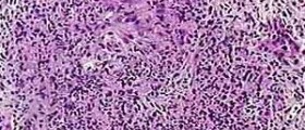
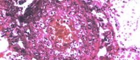
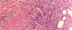
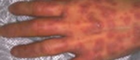
- Photo courtesy of Atlas of Pulmonary Pathology by Flickr: www.flickr.com/photos/30950973@N03/3734402837






Your thoughts on this
Loading...