Introduction to Painful Abdominal Masses
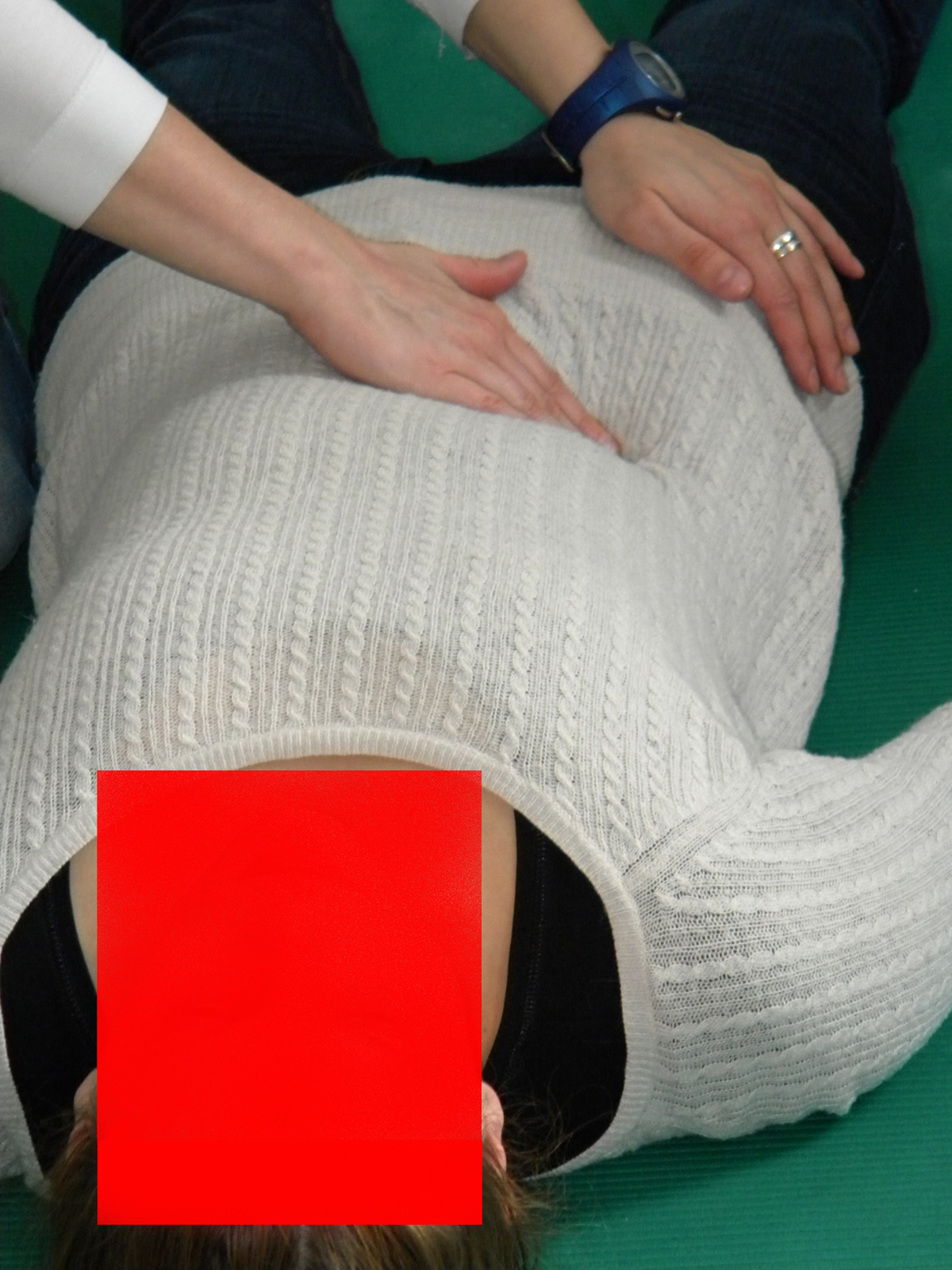
When a person has an abdominal mass it means that they have noticed some type of swelling in one part of the abdominal region, which is the belly and stomach region, generally speaking.
These masses can usually be detected easily by a doctor during regular physical examinations, and most of the time they develop slowly over time, so it might be hard to detect them yourself unless they become very painful.
The thing that helps the doctor give a proper diagnosis is when the patient tells them where exactly the pain is coming from. There are four general areas that make up the abdomen and there are specific organs and other body parts in each area. We divide the abdomen into the right-upper, left-upper, right-lower, and left-lower quadrants.
The center of the abdomen that is located right under the ribcage is referred to as the epigastric area and the periumbilical area is the part of the abdomen around the belly button.
- Discrete abdominal wall masses can be evaluated on the basis of their composition. Evaluation of the mass first includes confirmation that the mass is not a masslike process, such as a hernia. Next, the internal composition of the mass should be determined (eg, fat, fluid, solid, or fibrous composition). In a radiologic study of 365 patients who were referred to a sarcoma clinic for evaluation of an abdominal wall mass, the five most common masses in order of decreasing frequency were desmoid tumors (30% of the cohort), sarcomas of any type (20%), metastases (18%), lipomas (6%), and endometriomas (4%).
- Abdominal wall masses for which patients undergo imaging are more common in women than in men. In part, this is reflective of desmoid tumors being much more common in women and abdominal wall endometriosis being exclusive to women.
- The American College of Radiology (ACR) Appropriateness Criteria for palpable abdominal masses includes two variants: one set of criteria for palpable abdominal masses suspected to be intra-abdominal neoplasms, and a separate set of criteria for masses in the abdominal wall.
- The first step in evaluation of a potential abdominal wall mass is to determine whether it is a true mass or a hernia simulating a mass. If it is a discrete mass, the next step is to determine whether it predominantly contains fat, fluid, or soft tissue. Ultrasound, CT, and MRI are all useful for the differentiation of the tissue composition. At ultrasound, pure fat is usually hypoechoic, with an appearance similar to that of subcutaneous fat. Fat that is mixed with soft tissue, inflamed, or edematous is hyperechoic and attenuates sound.
- Abdominal wall fluid-filled masses or collections often have characteristic imaging appearances and are suggestive of certain clinical scenarios. These fluid collections can be characterized broadly as hematomas, fluid collections in postoperative patients, or cystic masses in patients without a history of surgery.
- Patients with abdominal wall masses that arise in areas of a prior surgical incision or biopsy can have a history that is suggestive of cancer, which may help to establish an accurate diagnosis. The primary consideration is abdominal wall metastasis in a patient who underwent prior abdominal surgery for cancer resection. Benign entities can also seed surgical incisions, and most abdominal wall endometriomas arise in women who have undergone cesarean delivery.
A diagnosis is created when taking into consideration the location of the mass, its firmness, texture, and other qualities that the doctor will be able to identify.
Causes
When a person has an abdominal aortic aneurysm they can feel a pulsating mass near the bellybutton and around that area.
Sometimes colon cancer will cause a mass to appear in the abdomen, but the location can depend from case to case.
Bowel obstruction, which is usually referred to as Crohn’s disease, can result in many tender and almost sausage-shaped masses in the abdomen.
Kidney, stomach, and liver cancer can also be another cause of masses in the abdomen.
Hydronephrosis is a condition in which the kidney is filled with fluid. This can lead to smooth and spongy masses in the flank area, which is the side of the body towards the back.
When a person is experiencing liver enlargement they can have masses right under the ribcage on either the left or right side.
Women with ovarian cysts can notice smooth and rubbery masses right about the pelvis, which is the lower abdominal area.
Pancreatic problems can lead to abdominal masses as well. When a person has a pancreatic abscess, for instance, they will have masses in the upper abdomen, but when they are suffering from pancreatic pseudocysts, the masses will also be in the upper region of the abdomen, but they will be lumpier in texture.
A ureteropelvic junction obstruction can cause a mass in the lower abdomen.






Your thoughts on this
Loading...