A Possible Bad Sign
Although children usually experience thigh pain due to overstraining of the muscles since they are involved in numerous physical activities daily, there are more serious causes of this condition. Namely, if a child feels pain in his or her thighs but the pain disappears in a day or two, this is a matter of no great concern.
However, if the pain remains persistent for a longer period of time, there might be a reason for getting worried. A pinched nerve may be behind this constant pain. Therefore, it is important to know the causes in order to take further measures and provide your child with an adequate treatment. Sometimes nerves get pressed by joints, bones, muscles or different tissue. This causes them to be dysfunctional resulting in pain, inflammation and discomfort.
This condition may be caused by numerous different factors. As mentioned above, mostly these problems stem from overuse. Nevertheless, there might be other causes as well. The child may have suffered a direct blow into his or her thighs, thus damaging a certain part of the tissue, resulting in pain and discomfort. Thus, examine your child well in order to establish the type of injury and take further steps regarding the possible necessity of a medical assistance.
Reasons Behind Thigh Pain in Children
First of all, children may experience pain due to the development of their organism. Growth of bones may cause pain in thighs.
Secondly, a bacterial infection may affect the bones of children usually aging from 3 to 12. This infection is mostly noticed upon a child's thigh bone, causing pain, irritation, redness and tenderness to the touch. Warmth may be felt upon touching the troublesome spot as well. If these symptoms are accompanied by swelling of the joints and fever with chills, one's joints may be infected by a more serious bacterial, viral or fungal disease called septic arthritis. Adults are just as prone to this as children are.
Thirdly, either benign or malign tumors may appear onto the surface of a thigh bone. While, the firstly mentioned are life-threatening and tend to cause pain, swelling and bruises, the latter are harmless unless they get infected. A type of bone cancer, osteosarcoma, may be behind the thigh pain experienced by a child. Usually affecting young boys, it causes pain, redness and swelling.
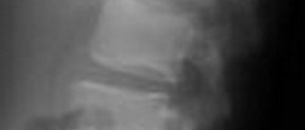
- Plain radiographs revealed a lobulated, cortically based lesion along the anterior aspect of the distal right femoral metadiaphysis with a relatively homogenous blastic appearance. Mature periosteal reaction was present as was thickening of the cortex. There was no obvious communication between the lesion and the femoral medullary cavity.
- A bone scan revealed an increased uptake in the lesion without evidence of other bony lesions. A chest computed tomography (CT) scan revealed no metastatic lesions. A magnetic resonance (MR) image revealed a 5.5 × 1.7 × 2 cm lobulated, cortically based mass with low signal intensity on T1 weighted sequences and intermediate to high signal intensity on T2 weighted sequences.
- Increased signal was seen in the underlying bone marrow on T2 weighted images. No significant soft tissue component was identified.
- The biopsy specimen showed proliferation of bland spindle to stellate cells without pleomorphism or mitoses. These focally condense into islands of relatively bland cartilage. Endochondral ossification or cartilage maturation was not recognized.
- The main tumor resection specimen shows similar histologic features. An osseous component, not obvious in the biopsy specimen, is present. The bony islands seem to be lamellar, which is often present in parosteal osteosarcoma. They are bland and condense from the spindle cell matrix.
- The patient initially underwent biopsy of the lesion at an outside institution via an anteromedial, vastus splitting approach. After the diagnosis of parosteal osteosarcoma was made, referral of the patient to the author's institution was made for definitive resection. Wide excision with limb salvage was planned, which involved resection of the anterior portion of the distal femur, with care taken to include the area of medullary involvement seen on the MR image. Neither the physis nor the joint were involved.
- There were no postoperative complications. Because the pathologic analysis revealed a well differentiated tumor with clear margins, and no dedifferentiated component was identified, chemotherapy was not recommended. The patient is alive and well with no sign of local recurrence or metastasis at 2 years after surgery.
Possible Treatment
Logically, the type of treatment greatly depends from the underlying cause. Therefore, after establishing a proper diagnosis, your child's doctor will prescribe the best possible therapy. If the cause is not a serious one, the pain will disappear on itself. Until this happens, hot compresses and resting are an excellent cure. More serious cases, however, require immediate medical intervention.









Your thoughts on this
Loading...