
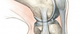
Knees are rather delicate structures which explains why they are easily damaged or injured. A variety of conditions affect the knees. Injuries are frequent among athletes especially those who participate in sports where the knees are excessively used such as running, basketball, football etc.
Injury of the Knee can be Diagnosed with MRI
MRI (magnetic resonance imaging) is a very potent technique that uses a magnetic field along with pulses of radio wave energy to visualize structures in human body. It is a highly powerful tool when it comes to diagnosing various knee problems. Practically each and every component of the knee area including muscles, ligaments, cartilage and bones can be properly visualized on MRI scans. Many times MRI of the knee will provide with more information compared to other imaging techniques such as X-ray or CT scan, especially when soft tissues in the area require thorough evaluation.
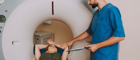
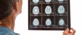
The injured knee or the knee affected by certain condition is placed inside the machine. After scanning the obtained images are evaluated. This procedure may easily confirm relatively tiny changes in the area and easily confirm the presence of severe damage or perhaps the presence of new growths affecting the bones or muscles. Digital images obtained during the procedure are stored in a computer and may be also available in the form of photographs and films. In case some structures that are next to each other cannot be distinguished enough, the patients will receive a contrast material. The contrast moves through blood vessels enhancing certain structures, making them clearer compared to surrounding tissues.
Doctors may order MRI of the knee in many situations. For example, the exam is indicated in case on abnormal finding on a knee X-ray or bone scan. Furthermore, MRI of the knee may easily confirm the presence of Baker's cyst, a build-up of joint fluid behind the knee. As a matter of fact MRI of the knee can easily confirm any fluid build-up in the knee area.
Furthermore, this technique is an excellent means of diagnosing infection of the knee joint, any kind of injury as well as benign or malignant tumors affecting the bones/muscles of the knee joint.
Also, MRI may be necessary once the injured knee is operated when doctors want to assess the success of the surgery.
Are any Preparations Necessary?
Most patients who undergo MRI of the knee are supposed to wear a hospital gown or clothing without metal fasteners which will make the exam much easier and will not interfere in the very procedure. All metal objects such as pens, pocket knives, hairpins, detachable dental appliances etc. must be removed. As a matter of fact, if patients have any metal implants or foreign metal objects inside their body they are not suitable candidates for MRI. What is more, MRI is not supposed to be performed in people who have had a pacemaker implanted.
The very test is done by lying on the table which slides into a large tunnel-like tube. Claustrophobic individuals may find it very hard during the test but since their head is not in the machine (only the legs) they may overcome the fear and undergo the entire procedure without psychological disturbance.
Patients who need to receive a contrast dye have an intravenous access prepared and once the doctor decided they are administered a dye. Contrast dyes are never given to patients allergic to any component of the dye. Also, allergy to certain medications may make patients prone to allergy to the dye itself which is also taken into consideration when deciding upon injection of these image-enhancing substances. And finally, contrast dyes are never administered in patients suffering from kidney disease or those who are on dialysis.
The exam does not last more than 60 minutes. All patients are closely monitored by medical staff from the next room.
Four to six hours prior to the exam patients should not eat or drink anything. Apart from that there is no specific preparation. Now, patients who are claustrophobic might be injected a medicine that will make them feel calm or a bit sleepy, removing the potential anxiety associated with being in the machine.
There is no pain associated with MRI exam. Patients are supposed to stay calm and not move at all so that obtained images would be of the best quality. The machine always produces loud thumping and humming noise which can be muffled with earplugs to certain extent. An intercom in the room is of great assistance when doctors want to give some advice to patients. This way they communicate without interrupting the exam by entering the room.
There is no recovery after the exam except for individuals who have received a sedative. They should never use vehicles after MRI but have someone to drive them home instead.
And finally, once the results become available patients come for a consultation with their doctor who makes a decision on further treatment.




_f_280x120.jpg)





Your thoughts on this
Loading...