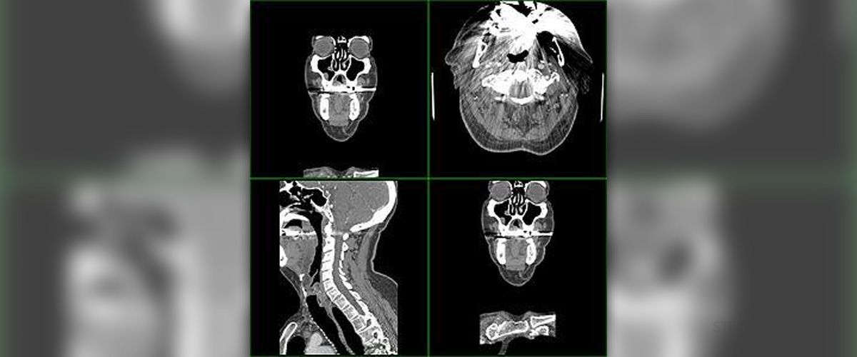
Sometimes, most commonly during the processes of medical diagnosis, a special type of scanning, called the CT scanning or CAT scanning is necessary. This procedure can detect many health problems and help the health practitioners manage to develop an adequate treatment plan.
Thus, if you have been directed to a CT scan during your medical treatment, the following lines will explain the process and help you go through it, knowing what exactly to expect.
What is a CT Scan of the Head?
CT scanning is a process which uses a special type of X-ray radiation in order to obtain images of one's inner body. When the scanning is carried out on one's head, the device focuses on collecting images of the brain, the skull and the sinuses. Additionally, CT scanning can produce images of the blood vessels in a person's head too.
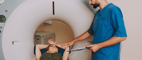
During this form of scanning, the patient needs to insert his/her head inside the circular machine. This machine then circles around the head area, exposing it to radiation from various angles in order to obtain a final, 3D image, processed by a computer connected with the scanner. Therefore, the images can be both 2D, resembling a normal X-ray scan, or 3D, showing the images of the head in many details, broadening the perspective.
The whole process of scanning is performed by a radiologist specialist, being a qualified member of the medical staff.
As far as the reasons behind CT scanning of the head area are concerned, these are mainly connected to diagnosing certain medical conditions according to the information obtained from the images. So, a CT scan can detect conditions like hydrocephalus, brain swelling, inflammation, bleeding and injuries. Also, it can locate and define abscesses, as well as cysts and tumors, or even reveal some birth defects, if these are present in the skull area or the brain.
Furthermore, CT scanning diagnoses the health status of the pituitary gland, the pineal gland and the sinuses, along with the blood vessels located in the head. Therefore, it can define the causes of headaches, fatigue or some mental problems.
Before a person undergoes a CT scan procedure, he/she is not allowed to eat or drink anything for a couple of hours, especially in cases when a special substance called the contrast dye is injected in the blood vessels, for the purpose of scanning or when sedatives need to be administered prior to the procedure.
The patient needs to remain still during the scanning. He/she is allowed to communicate with the doctor or the health practitioner through the intercom devices installed in most CT scans. The process is not painful and it does not lead to any side effects. Yet, the patient may hear the buzzing noise while the machine is working and the area inside the device may seem cold and uncomfortable. Nevertheless, the patients are advised to bear with the scanning process, since the benefits of such procedure are countless and crucial for the rest of their treatment.
Once the images are obtained, the radiologist will examine them and assess the results, reporting these to your doctor, who will let you know about your health condition. Most commonly, the results are complete over the course of several days.
You should bear in mind that CT scanning delivers more X-ray radiation than a simple X-ray scan. Moreover, this radiation is stronger than the regular one, even though the radiologists are bound to use minimal amounts for obtaining the required results.
Cranial and Extracranial Scan
Cranial scans and extracranial scans are used for different purposes, during the diagnosis of different forms of health conditions.
Thus, in cases of acute stroke, CT scanning can be done within 48 hours after the incident, ruling out any internal bleeding or detecting it timely. Also, this form of CT scanning can be used for detecting or ruling out primary intracerebral hemorrhage, a week after the onset of this condition. Thrombosis or endarterectomy patients can also be suitable for CT scanning, during the diagnosis and monitoring of their conditions.
Also, cranial CT scans can be used in patients who have problems with transient ischemic attacks, acute subarachnoid hemorrhage, acute head injury, space-occupying lesions, epilepsy, personality changes, dizziness, coordination issues or any appearance of numbness or tingling affecting the area.
Furthermore, intracranial infections may need this form of scanning and it can also be used in cases of headaches, brain herniation, head injuries etc.
As for the extracranial scanning, it is directed towards the process of diagnosing, treating and assessing conditions such as middle or inner ear health problems and balance complications which go with it, even though MRI scans are a better alternative in these cases.
This form of scanning is useful in cases of sinus disease, congenital anomalies, orbital lesions, fractures of the bones in the head area and all other cases where MRI scanning is considered unsuitable.


-Test-And-What-Do-The-Results-Mean_f_280x120.jpg)






Your thoughts on this
Loading...