Lumbar spine and MRI
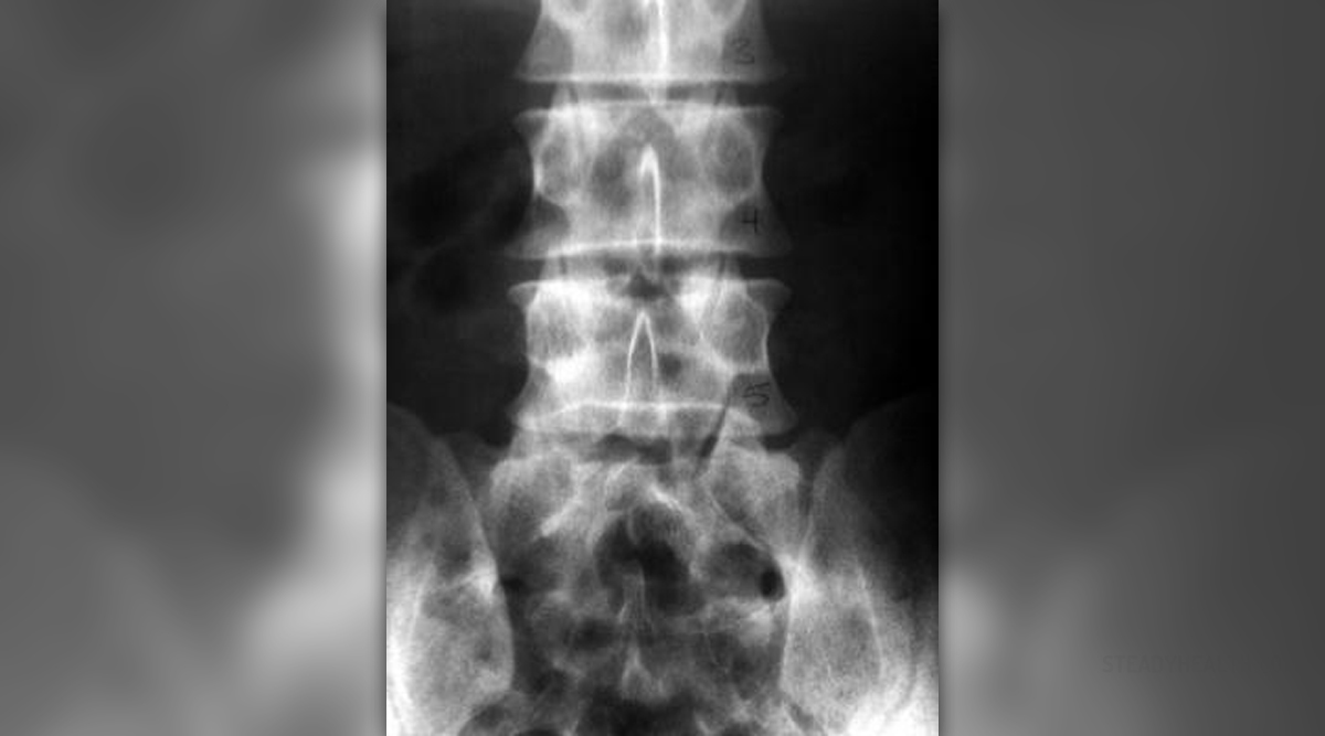
The lumbar spine is the part of the spine in the lower back. Five vertebrae called L1, L2, L3, L4 and L5 comprise the lumbar part of the spine. The best way to see a detailed picture of the lumbar spine is to use a lumbar magnetic resonance imaging – MRI. MRI is a non-invasive procedure that uses radio waves and powerful magnets. The MRI has a magnet whose magnetic field is even ten thousand times greater that the earth’s magnetic field. This magnetic filed forces the hydrogen atoms to gather together or line up in a certain way and when the radio waves reach these lined-up hydrogen atoms, they tend to bounce back. At that point, the computer records the signal.
Performance of the test
The patient should wear a hospital gown without any metals on him. Then, he/she lies on a table that slowly slides into the middle of the MRI machine. Sometimes, the patient is given a dye before the test so that the specialists can sea some thing more clearly. It is given through a vein in the arm. The exam usually lasts about an hour or more, but it depends on the equipment.
The patient should not eat or drink anything for four or six hours before the exam. The doctor should be informed if the person is a diabetic or is a claustrophobic. Furthermore, the people who have cardiac pacemakers cannot have MRI. Moreover, metallic objects usually interfere with MRI and when a person has something metallic in the body, as well as on the body, he/she cannot have MRI. The MRI test is not painful, but it can cause anxiety since one has to stay lying inside the MRI machine for an hour or longer.
Lumbar spine MRI
The lumbar spine MRI is ordered when certain symptoms occur, such as injury to the lumbar spine, birth defects of the lumbar spine and infection that includes the lumbar spine. Furthermore, in case when scoliosis or multiple sclerosis is present, lumbar spine MRI is ordered also. Moreover, when the pain or numbness occurs in the low back, this test is also recommended. Syringomyelia and tumor or cancer in the spine also require lumbar spine MRI.
When MRI shows that the lumbar spine and surrounding nerves are normal in appearance, it is considered to be normal results. Abnormal results usually point out certain disease, such as spinal tumor, spinal cancer, disk degeneration, Cauda equina syndrome, diskitis and herniated disk, as well as osteoporosis, spinal stenosis and syringomyelia.


_f_280x120.jpg)


_f_280x120.jpg)


Your thoughts on this
Loading...