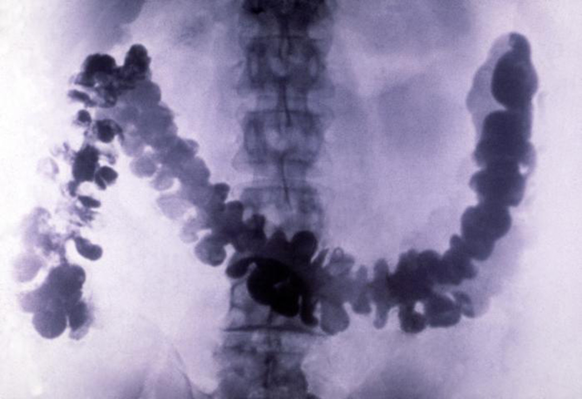
Barium Enema
Barium enema also known as the double-contrast barium enema is an X-ray test which is used to visualize gastrointestinal tract. It uses white liquid barium which is injected through a catheter through the anus and into the rectum and from there it fills the large intestine. The purpose of administration of barium is better visualization of the intestines which cannot be seen properly on plane X-ray of the abdomen. Barium provides proper contrast to surrounding structures such as solid organs and this way the large intestine can be seen clearly and with more detail. Barium enema helps in identification of certain intestinal abnormalities such as diverticulitis, polyps, abscesses, dilatation, cancers and so on.
Risk of Barium Enema
Filling of the intestine with barium cases distension of the intestine which may be rather unpleasant and only small number of patients actually complain about the pain during the procedure. Another risk is the actual exposure to X rays. The exposure is limited and today's techniques prevent excessive exposure of the surrounding tissue to X-rays.
The risk of exposure is high for pregnant women and they need to report the pregnancy before the exam. X-rays may cause serious damage to the fetus especially if this examination is performed during organogenesis. Pregnant women are, in general, supposed to avoid this examination.
This examination is never performed if there is even the slightest suspicion that perforation of the intestine has occurred.
One of the potential complications includes abscesses and peritonitis. They occur if during the procedure the intestine is accidentally perforated by the catheter.
The Very Procedure
Since the presence of the feces inside the intestines may interfere in visualization of these structures, patients are due to clean the intestine prior the procedure. This can be perfectly achieved by a clear liquid diet and cleansing enemas. They will empty the colon and provide with better adhesion of the contrast. Apart from a clear liquid diet and colon cleansing some patients require additional medication to cleanse the colon properly.
External clothing and all the metal objects must be removed. Metal can also give certain presentation and interfere in adequate visualization of the colon.
The colon is filled with white, liquid barium via catheter. Then an X-ray machine is drawn and placed in front of the patient. The X-ray film is behind the patient. Images of the colon are obtained after the colon filled with barium is exposed to X-rays. Fluoroscope is a machine used by radiologist to visualize the colon and eventual abnormalities. All the information is recorded on the film. The barium is then drained and the colon is filled with the air. Another set of X-rays is applied. This is typically performed in double contrast barium enema. And finally, the air will spontaneously drain from the colon which is the end of the examination.











Your thoughts on this
Loading...