
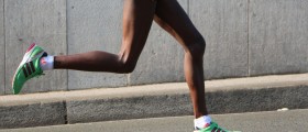
Running is a sport which is quite hard on the body. However, the passion for running that many people experience helps them overcome such dangerous trait of this activity. Nevertheless, running injuries are a common scenario for all runners. Since these injuries appear frequently once you are a dedicated runner, read the lines below to learn more about these phenomena and possible ways of treating them.
The human foot consists of 26 main bones, which can further be divided into 3 different regions. These are the forefoot, the midfoot and hindfoot. Each and every bone has its own purpose, making our feet as useful as they are.Running Injuries
There are several injuries to the foot which are most commonly seen in runners. The first one is plantar fasciitis, manifesting through pain felt in the heel area due to inflammation of the thick ligament at the base of the foot. This pain gets worse when the person is running and, if left untreated, evolves into a heel spur.
Secondly, overpronation is yet another condition which is not unknown to all those who practice running on a daily basis. When the foot has moved through the gait cycle for too many times, pronation occurs, interfering with the proper mechanics of the gait cycle. Fortunately, this can be prevented by specially designed shoes.
Finally, one of the most common conditions runners suffer from is arch pain, which is, basically, an inflammation appearing at the arch of one's foot. This leads to a burning sensation, felt in the area. Again, timely treatment with adaptive footwear and special insoles is known to help.
Difficulty of Diagnosing
In order not to experience any problems regarding the diagnosis of foot injuries, doctors have Ottawa Foot and Ankle Rules, which help them find out what people need radiographic evaluation.
Basically, those who cannot carry any weight for at least 4 steps, once they have injured their feet, need to undergo radiographic evaluation. The same goes for people who show or tell that their posterior aspect of the medial and lateral malleoli, over the navicular or over the base of the fifth metatarsal area is tender.
Diagnosis of foot injury patients is usually very precise, having a 0.3% sensitivity rate in adults and 100% sensitivity rate in children. Nevertheless, the specificity approaches about 30 to 40%.Diagnosing the Most Common Foot and Ankle Injuries
As far as turf toe is concerned, the diagnosis will involve plain radiographs. This kind of approach will possibly reveal any small avulsion fractures appearing on the plantar metatarsal head. If sesamoiditis is the case, stress radiographs, axial silhouette views, bone and CT scans can all aid the process of diagnosis. Sometimes, radiographic examination of the collateral area of the foot may be necessary, along with some other forms of testing, based on the signs the patient is showing.
Plain radiographs are usually not needed in cases of severe disease. Yet, if resting fails to remove or reduce the symptoms of this condition, this approach may be helpful in the process of ruling out breakages or tumors.
In cases of posterior tibial tendinitis, the level of flat foot condition needs to be determined and this is done through a weight-bearing plain radiograph. Ultrasound or MRI scanning may also be necessary for obtaining a better overview of the condition.
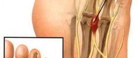
If a patient is bothered by peroneal tendon subluxation or dislocation, no special treatment is necessary since this condition disappears on its own in most cases. Yet, if tendinitis appears in this area plain radiographs, US imaging or MRI scans may be useful in the diagnostic process. Radiographs are also known to be helpful with FHL tenosynovitis, Jones fracture and Morton neuroma, especially in the process of determining the size of the neuroma, as far as the latter is concerned.
Moving on to stress fractures, these are usually diagnosed through plain radiography. Yet, this approach may not be capable of noticing any changes appearing during the first 2 or 3 weeks after the appearance of the symptoms. Thus, only 50% of stress fractures are observed this way and MRI scans provide additional support during the first 2 weeks.
Lisfranc fracture dislocation requires careful evaluation of weight-bearing foot radiographs. Additionally, MRI scans and US imaging may be necessary during the process of diagnosing this condition.
When to Return to Running?
Once a foot injury heals completely, the person needs to be very careful before returning to his/her previous physical activities. Namely, he/she should practice before any serious indulgence in his/her activity, making sure that the whole process can be carried out without the presence of pain. The affected, previously injured, limb needs to possess at least 90% of the healthy limb's strength in order for the foot to be considered healed. Thus, patients should be very careful and return to their previous activities gradually and carefully.
All in all, there are many different types of foot injuries which can appear due to running. Thus, be careful and have your foot problem diagnosed and treated timely.






Your thoughts on this
Loading...