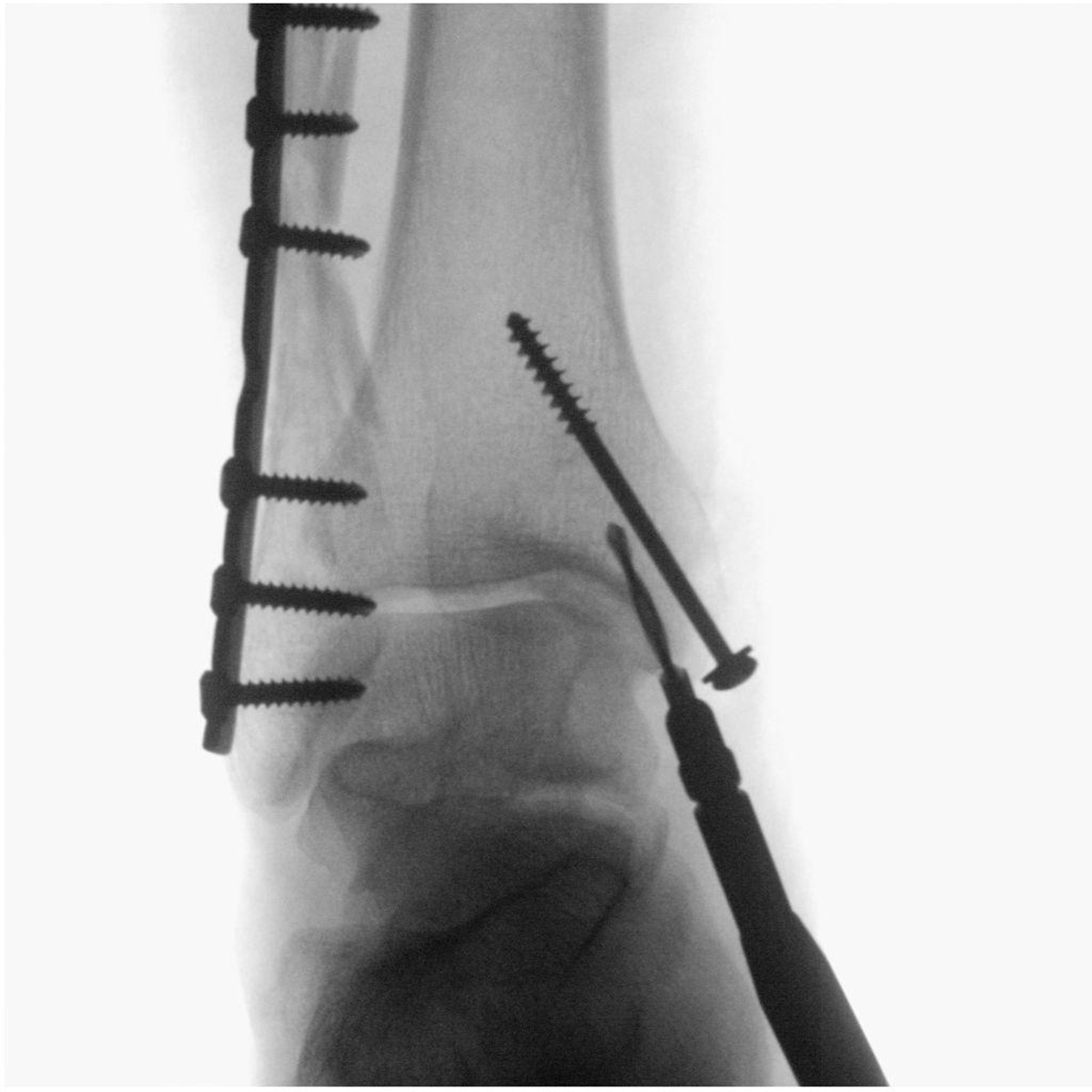
What is fluoroscopy?
Fluoroscopy is a medical imaging technique performed mainly for diagnostic purposes. It uses a device called fluoroscope to obtain real-time images of a patient’s internal structures.
The simplest form of fluoroscope consists of a source of X-rays and a fluorescent panel. The patient is asked to stand between the two in order to obtain images. Modern fluoroscopes also consist of a CCD video camera and an image intensifier.
One of the major concerns regarding the use of fluoroscopy is the amount of radiation a patient receives. The X-rays are used in form of ionizing radiation and today the amount of radiation is relatively low. However, because the procedure can be quite lengthy, the total amount of radiation can be significant. Because of this, it is sometimes difficult to establish whether this imaging technique provides more harm than actual good. Fortunately, today the risks are significantly lower than in the past, mostly because digitalized images and flat screen detectors, which reduce the risks.
Fluoroscopy equipment
The first fluoroscopes were made of a fluorescent screen and a source of X-rays. The patient would stand or sit between these two parts and as the X-rays pass his or her body they cast a shadow on the screen or panel. The X-rays interact with the atoms in the fluorescent screen through the photoelectric effect. The images were at first very dim, which is why radiologists needed to read the images in a darkened room or using special goggles. With the invention of image intensifiers it was made possible to observe the images at normal light too. Video cameras, and later, CCD cameras, made it possible to store the images electronically.
Flat-panel detectors improved fluoroscopy even more. They offer increased sensitivity to X-rays and thus reduce the radiation dose that a patient receives.
Procedures
There are many medical procedures that involve the use of a fluoroscope. In the gastrointestinal tract, fluoroscopy is used in barium enemas, swallowing and meals, in defecating proctograms and enteroclysis. This technique is also used in orthopedic surgery. Angiography of the leg, blood vessels and heart uses fluoroscopy, and so does urology, especially retrograde pyelography. Fluoroscopy is an important aid in surgical procedures for placing pacemakers, defibrillator and other devices that help the heart function properly. It is also used in placement of eating tubes, especially if previous attempts without this technique have failed, as well as in placement of central catheters, such as PICC.








Your thoughts on this
Loading...