Discovery of X-rays
Wilhelm Conrad Roentgen discovered X-rays in 1895 when his team of scientists were studying the pathways of electric currents passing through a vacuum tube. By accident, they noticed a bright light when the current went through. Roentgen took an X-ray photograph of his wife’s hand and realized it showed her bones and wedding ring. The discovery sparked great interest in the scientific community, and within only a few months, the X-rays were being used both in Europe and the US. Their initial aim was to reveal internal organs and subsequently treat and prevent diseases. As the rays were obscure the scientists named them X-rays, and the name stuck much to Roentgen’s objection. The rays are called the Roentgen rays in many German speaking countries. It was later discovered that X-rays are a form of invisible, high frequency electromagnetic radiation. They have an abundance of energy and get easily through bones and tissue. The amount of energy that does not pass through the organs or the tissue is stored inside the body. The residual energy subsequently leads to problems caused by radiation. There are various amounts of energy used for different purposes, with cancer being one of the diseases that requires the most radiation. A type of photographic film is utilized with X-rays and once they are changed into light the film becomes darker. The bones being a dense tissue appear lighter in X-rays as they have a hard time passing through. On the other hand, if the tissue is relatively solute the X-rays are easily detected. In addition, a single X-ray provides plenty of information about what is going on in internal organs, such as if there are changes on the lungs or if there are any bones which are fractured. Further, there are various techniques which aid the X-rays in showing blood vessels or the movement of bowel. X-rays are predecessors of CT scans, which are able to generate a serious of pictures and capture the functioning of an organ over a consecutive period of time. Further, taking of an X-Ray image is a fairly simple procedure. An individual who needs scanning is positioned between a machine and a screen which generate the picture. Anything that a person wears on their body needs to be removed in order to avoid any confusion. The patient needs to be completely motionless so that there is no shading in the picture. It should be noted that the procedure is painless and there are no short term side effects. A technician usually takes the images, but they are sent to a radiologist who gives a formal diagnosis. In some simple cases the interpretation is available right after the taking of the images, such as at a dentist’s office. Further, the X-rays emit radiation so in the long run they can lean to cancer. On the other hand, the radiation produced by X-rays is utilized in cancer treatments. In modern medicine, a very small amount of radiation is needed to produce good quality images so individuals who need to be scanned need not worry about cancer development as a result of exposure. However, medical care professionals who administer X-rays would be exposed to a substantial amount of radiation so in order to avoid it they usually either leave the room or stand behind a screen while the images are being taken. In addition, amount of radiation exposure from X-rays is enough to lead to problems in a fetus so pregnant women are advised against scanning as much as possible. Most technicians or doctors administering X-rays ask all women patients if they might be pregnant. Minor scanning, such as that of teeth, would be avoided by most dentists if the patient were pregnant.
What Are X-Rays Used for?
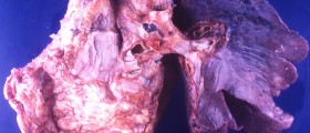
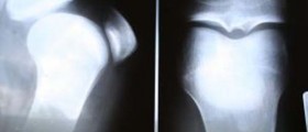
X-Rays are utilized to examine the chest, or in particular look for any anomalies in the lungs, heart, or major arteries. Pneumonia is a condition easily diagnosed with the use of X-Rays, and so are respiratory illnesses and lung cancer. Disorders of the cardiovascular system are also detectable, while images of the bones come out the clearest. Bone illnesses that are diagnosed through the use of X-Rays are cancers, osteomyelitis, teeth problems as well as bone fractures.
Potential Risks Involved in X-rays
The risk of developing cancer from exposure to radiation via X-Rays exists, but it is fairly low. One dosage necessary to perform a scanning of the chest, for instance, produces the possibility 1 in 1 million that the individual will develop cancer. The risk is slightly higher for head imaging, but is still minor. When it comes to breast and abdomen X-Rays, they generate yet a higher risk, with the highest being produced by barium swallowing. It should be noted that every individual has a 1 in 3 possibility to develop cancer at some point in life.


-Test-And-What-Do-The-Results-Mean_f_280x120.jpg)


Your thoughts on this
Loading...