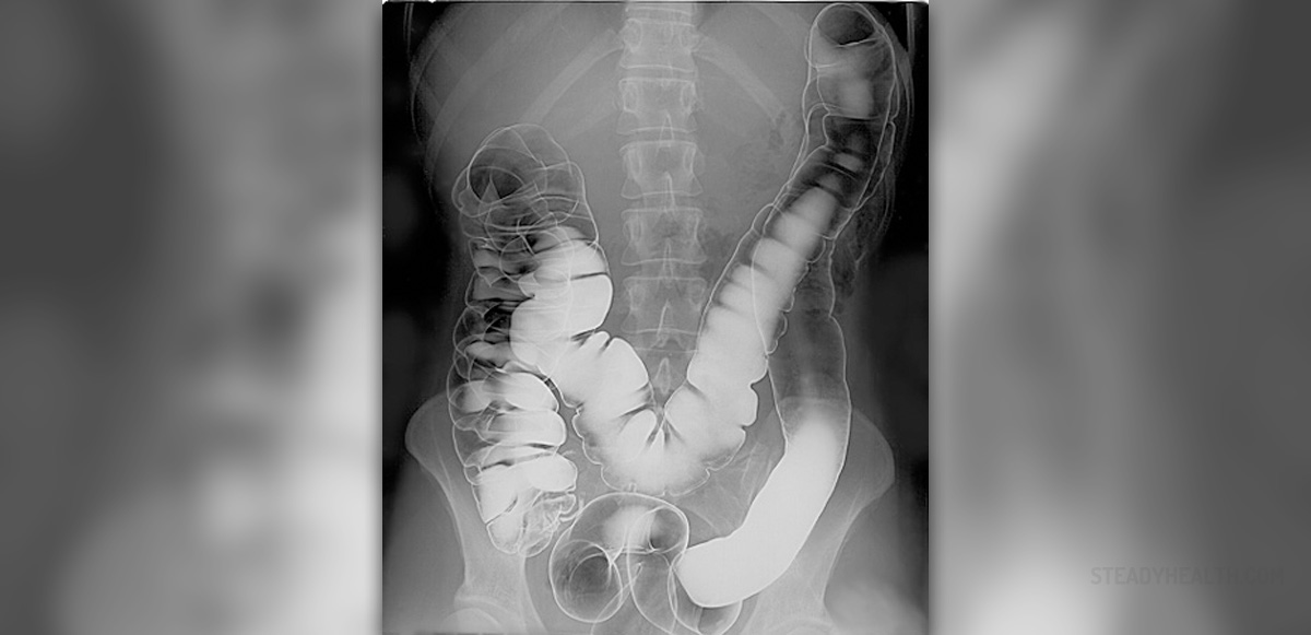
Barium enema is an imaging procedure performed as an outpatient procedure. Its goal is to use contrast to explore the large intestine using a contrast in the bowel created by barium, which is observed through a regular x-ray.
In this test, a particular portion of the bowel is examined to determine any abnormalities or diseases in the lower gastrointestinal tract.
About barium enema
Barium enema test is performed to examine the large intestine. This particular part of the gastrointestinal tract is filled with air, so normal x-ray goes right through it, producing a plain picture that cannot be used for diagnostic purposes. Barium enema uses the contrast that is created after the barium is introduced, so the image can show the exact state of the bowel.
This procedure is usually done by a radiotherapist at the radiology department of a hospital. It is an outpatient procedure, which means the patient does not have to stay at the hospital. It is a simple test, but there are some preparatory steps that need to be done prior to a barium enema.
Preparation for barium enema
The most important step to do before barium enema is to empty the colon. If the colon is not empty, the image will be useful and there is a risk of getting the wrong results. Radiologist provides tips as to how to achieve an empty colon, and he or she also instructs the patient not to eat or drink anything four hours before the procedure.
The patient also needs to see a doctor who will examine the medical history and possible allergies.
Barium enema procedure
There are two types of barium enema procedure- single contrast and double contrast. In single contrast, an x-ray is taken directly after the barium solution is introduced. In double contrast procedure, the barium is filled in the bowel and then emptied. After this, the portion of the bowel is externally filled with air.
Unlike single contrast, double contrast barium enema provides a detailed picture of the intestinal lining.
During barium enema, the patient is asked to lie flat on the back on an x-ray table. A normal x-ray image is taken before the introduction of barium. After this, with the patient lying on the side, the doctor gradually fills the bowel with barium through the rectum, using an enema tube attached to a bag with barium. This may cause a sensation of fullness, mild discomfort and an urge to defecate, which is normal. The doctor follows the flow of barium through a fluorescent screen and then takes x-ray images from different angles. After this, the enema is removed and the procedure is completed.
The procedure lasts from 30 to 60 minutes. The patient may experience whitish stools in the next few days. Doctors usually recommend drinking a lot of water or juice to flush out the barium.








Your thoughts on this
Loading...