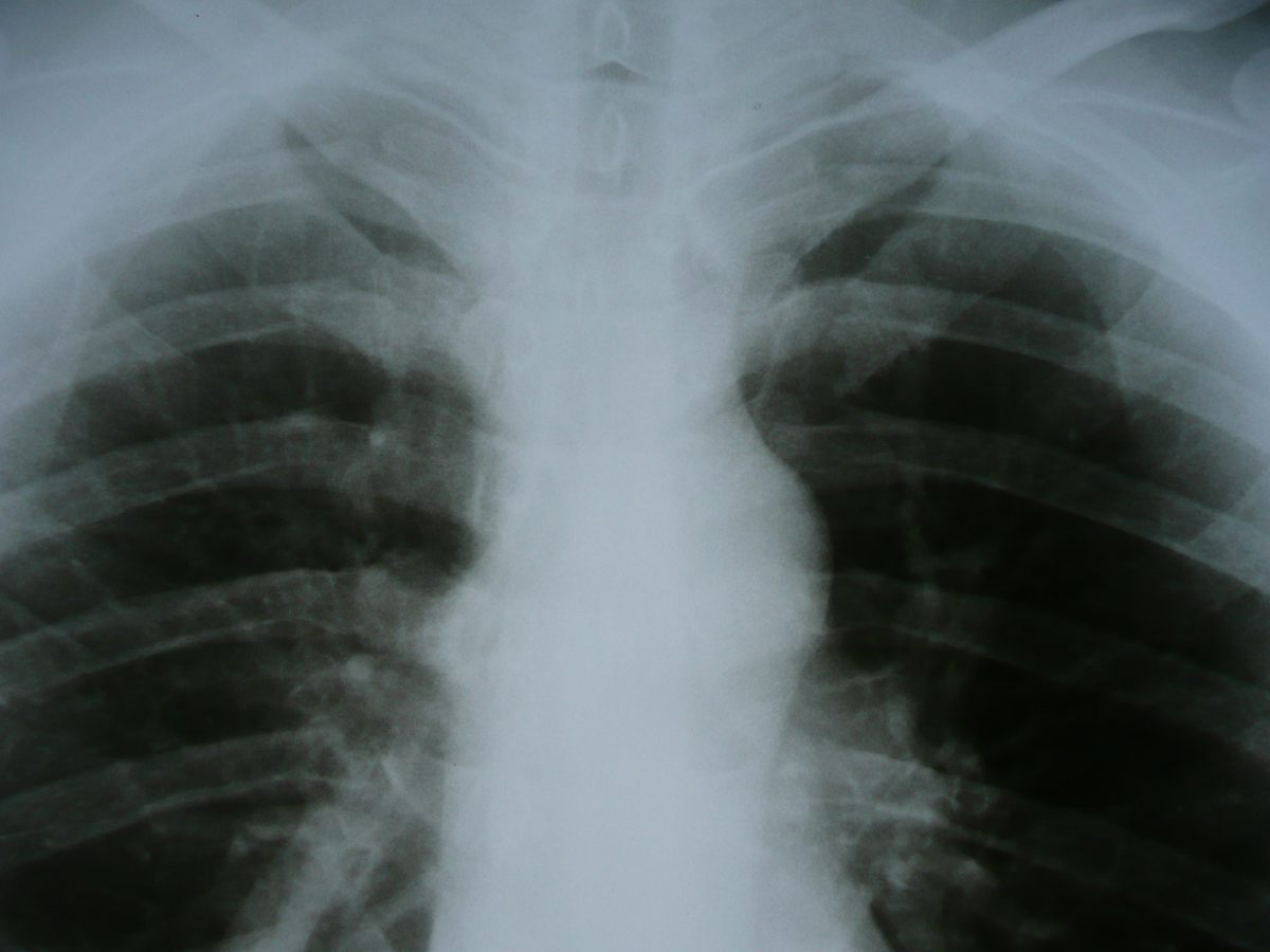
Today, many health problems are diagnosed through radiology imaging. Moreover, various diseases can even be treated through radiology.
Thus, if you have been told to undergo a radiology imaging procedure, read the lines below and learn many useful things about this procedure, its purpose, its performance and many other factors.
What is Radiology?
Basically, as it was mentioned above, radiology is a medical procedure involving using special types of imaging in order to diagnose and treat specific disease found in the body of the patient. More than a single type of radiation used for these purposes exists. Namely, X-ray radiography, ultrasound, CT scanning, positron emission tomography or PET scanning as well as MRI or magnetic resonance scanning, all are possible tools of radiography.
Regardless of the type of scanning one undergoes once he/she visits the radiology department, the whole procedure is carried out by radiographers or radiologic technologists.
Acquisition of Radiological Images
The type of acquisition of radiological images, depends on the method used for obtaining them. Thus, first of all, one may need to undergo projectional radiography. Here, the X-rays are produced and emitted through the part of the patient's body while a capturing converts them into images. Most X-ray scanners of today still use the classical film imaging. Here, the rays which pass through the body are stored on the film which is kept in a dark environment.
However, today, there are new methods getting more and more popular in the world of medicine. One of these is digital radiography, where the images are instantly transferred onto a computer screen, possibly being analyzed and interpreted by the computer software itself.
Speaking of X-ray imaging, there are other types of this radiology. Fluoroscopy, being one of these consists of a fluorescent screen connected to a short-circuit television set. Here, the patient's scanned images are displayed live, during the imaging process itself. Yet, beforehand, he/she needs to take special radiocontrast agents by swallowing them or having them injected.
Intervential radiology is yet another type of this medical procedure, being specifically designed for diagnosing or treating certain diseases through using minimally invasive techniques. The images received through this process serve as guidance during actual surgical procedures such as the treatment of renal artery stenosis, inferior vena kava filter placement, gastrostomy tube placements and many others. Basically, once the surgeon has the images, he/she can use minimally intrusive procedures in order to carry the surgery out, greatly decreasing any chances of infections affecting the patient afterwards.
Moving on to CT scanning, this technology uses X-rays too, in concordance with computer software. Here, X-ray devices rotate around the patient, taking images and allowing the computer to create a cross-sectional image, also called tomogram. Here, radiocontrast agents can be used too, depending on the purpose of the scanning. Even though CT scanning exposed a patient to more X-ray radiation than a regular X-ray scan, it also provides better images, increasing the detecting capabilities of the procedure.
Some types of CT scans can even provide 3D images, helping doctors to diagnose and treat specific conditions significantly.
Finally, the ultrasound is a form of radiology using high-frequency sound waves in order to obtain images of the soft tissue in our body, appearing live during the scanning procedure. Here, there is no ionizing radiation. Therefore, this procedure is much safer than the other ones mentioned above. Yet, ultrasound imaging needs to be carried out by a skilled professional since this increases the quality of the received images incomparably. Also, the very body of the scanned patient plays an important role when it comes to the quality of the images.
Due to ultrasound's inability to go through air, it cannot display bones, lungs, bowels and other such structures. This form of scanning is mostly used for monitoring pregnancies and locating any cases of hemorrhage or other problems in the body.
Magnetic Resonance Imaging
Magnetic fields can also be used in radiography. An MRI scanning device functions on the principle which involves aligning the atomic nuclei in the body tissues and then exposing them to a radio signal which disturbs their rotation. Through the differences between the two scanned states, the images are created.
Today, MRI devices can create 3D images to, with the help of computers. Therefore, MRI imaging is a very useful procedure for muskuloskeletal radiology and neuroradiology.
In the US, in order to become a radiologist, a person needs to complete the necessary undergraduate education, followed by 4 years of medical school, a single year of internship and 4 years of residency training. Even when all this is behind you, you may desire to take one or two additional years of specialization, taking into consideration that the competition for this line of work is quite fierce, according to various researches.
All in all, radiology is an umbrella term encompassing many different forms of imaging and scanning performed in order to diagnose and treat specific health issues. Depending on the disease or other forms of health problems themselves, different types of scanning are used.
Radiology is an excellent method for maintaining your health and having the causes behind your illnesses identified. Thus, take advantage of this aspect of medical support whenever you need it.









Your thoughts on this
Loading...