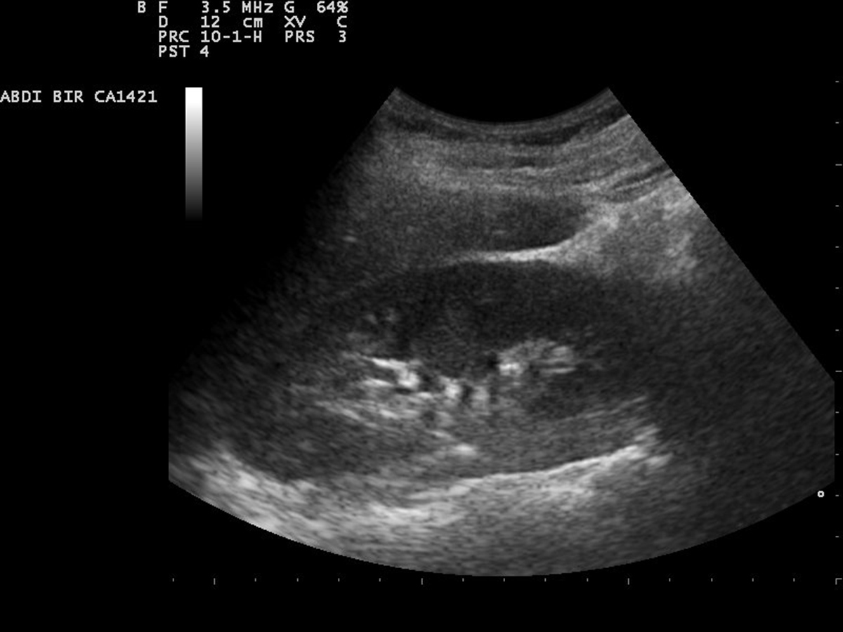
A kidney (renal) ultrasound is a simple, painless, noninvasive, and very accurate exam that gives perfect insight in the very structure of the kidneys. A kidney ultrasound is performed to help doctors evaluate potential anatomical abnormalities of the organ and confirm or rule out certain medical conditions.
Ultrasound of the Kidneys
The kidneys are located in the abdomen, in the retroperitoneal area. Their primary role is to excrete waste products, excess water and electrolytes from the body. The urine synthesized in the kidneys is pushed down the ureters, and then enters the bladder (the perfect reservoir) and eventually gets excreted through the urethra.
The functioning of the kidneys is affected in many different medical conditions. Some of these medical conditions cause changes that might be confirmed with available imaging methods (e.g. ultrasound, CT or MRI), while others trigger serious symptoms and can be only confirmed when tissues of the kidney are biopsied and microscopically evaluated.
A renal ultrasound is a powerful tool used in cases when patients are suspected to suffer from certain conditions or have suffered an injury to the organ. This kind of ultrasound can also visualize congenital abnormalities, confirm the presence of certain types of kidney stones, and assess other types of blockage. A renal ultrasound is also very helpful when it comes to assessing complications of a urinary tract infection that has reached the kidneys, or damage associated with cysts and tumors.
Now, as far as kidney stones are concerned, there are several types of kidney stones. Ultrasound can be efficient in visualizing clear uric acid stones and obstructions caused by any type of kidney stones. Most scientist agree on the fact that ultrasound simply is not sufficient enough to detect very small kidney stones. However, ultrasound scan remains a very useful first diagnostic step in emergency rooms.
Apart from ultrasound, a standard X-ray of the kidneys may also reveal some kidney stones, and calcium kidney stones in particular. This imaging technique is also helpful when it comes to diagnosing cystine crystals. The best diagnostic tool for kidney stones of any type is most certainly a helical computed tomography scan.
How is a Renal Ultrasound Done?
So, you are about to undergo a kidney ultrasound? As for preparation there is practically none, which means that this type of medical exam can be easily performed at any time. However, in order for the kidneys to be visualized with greater ease, patients should not eat or drink anything several hours before the test, if they are aware they will be having a kidney ultrasound in advance.
A renal ultrasound is performed in the radiology department of the local hospital. It is also possible to undergo examination in a radiology center. The clothes you wear on the upper part of the body should be removed, because the probe comes into direct contact with the skin. The presence of clothes interferes with the exam. The person lies down on a table, usually on his or her side so that the back remains approachable to the radiologist. The room is supposed to be dark. Reduced light allows better insight in discrepancy between white and black shades on the computer screen.
The inspected area is covered with a clear, warm, water-based gel. The gel does not allow any air to come between the patient's skin and the probe (which is called a transducer). Once the kidney ultrasound exam starts, the probe sends ultrasound waves to the patient's body. These waves enter the body, and some are absorbed, while others are refracted and return to the probe. The waves are collected and finally form the images on the computer screen.
By moving the probe and sending waves from different angles, the radiologist obtains a clear image of the kidneys and gains better insight into their structural changes (if there are any). The entire procedure does not last longer than 30 minutes and probably less, and a kidney ultrasound is completely painless. Any pain associated with kidney ultrasounds is only reported in individuals who were already experiencing discomfort or pain in the flank area prior to the exam, and results from the fact that the transducer is pressed down slightly.
Ultrasound is generally a highly safe procedure associated with no radiation. Because of that, it is indicated and recommended in people of all ages, and both genders and is safe for pregnant women. What is more, the exam does not last long so it can be utilized when quick evaluation is necessary which is the case with many emergency conditions.
In case of acute renal colic, when there is a chance the person is suffering from kidney stones, doctors take into consideration several factors. The first one is the patient's clinical presentation. The second is urinalysis (the results of a urine test) and finally, there are imaging techniques, ultrasound being the first procedure of choice.
The exam is supposed to rapidly evaluate the patient and give answers to several questions. For example, the ultrasound may easily confirm or rule out hydronephrosis (swelling of one kidney due to a backup of urine), the presence of fluid around the kidneys, distension of the renal pelvis, and the presence of kidney stones which can be identified with ultrasound.
After the initial assessment, patients may be asked to undergo additional exams such as IVP (intravenous pyelogram) or a CT scan. Both of these are highly efficient in confirming the presence of kidney stones when these cannot be adequately evaluated with ultrasound.
All in all, a renal ultrasound is a very useful exam which allows doctors to quickly evaluate the condition of kidneys without exposing patients to radiation or using IV contract. The test can be repeated without any harm. Still, it cannot reveal every type of kidney stones, therefore patients usually require additional exams.


Your thoughts on this
Loading...