Introduction to Valgus Knee
Valgus knee is a joint deformity which features with the outward angulation of the knee. The deformity is basically caused by the rheumatoid arthritis, post-traumatic arthritis, osteoarthritis or metabolic bone disease. The valgus knee comprises two components, a bone loss accompanied by metaphyseal remodeling and a soft-tissue contracture which consists of tight lateral structures.
In valgus knee a line which starts in the middle of the hip and runs to the middle of the ankle does not go through the knee but it runs through the outside half of the knee. This puts excessive load on this particular part of the knee and causes wear of its structures. One solution for this wear is a procedure called osteotomy. The principle of this procedure is to realign the leg. This way the line that connects the middle of the hip and the middle of the ankle will actually run through the middle of the knee and the load will be divided and carried by both halves of the knee equally.
The Surgery for Valgus Knee
The goal of the surgery is to restore the alignment of the knee, the joint line, the stability of the joint and its range of motions.
Prior the surgery the doctor performs multiple radiographs of the affected knee which will help him/ her evaluate the type of valgus knee and plan the surgery. After they are admitted to the hospital patients are operated the very day. They are forbidden to eat or drink anything for approximately 6 hours prior the surgery.
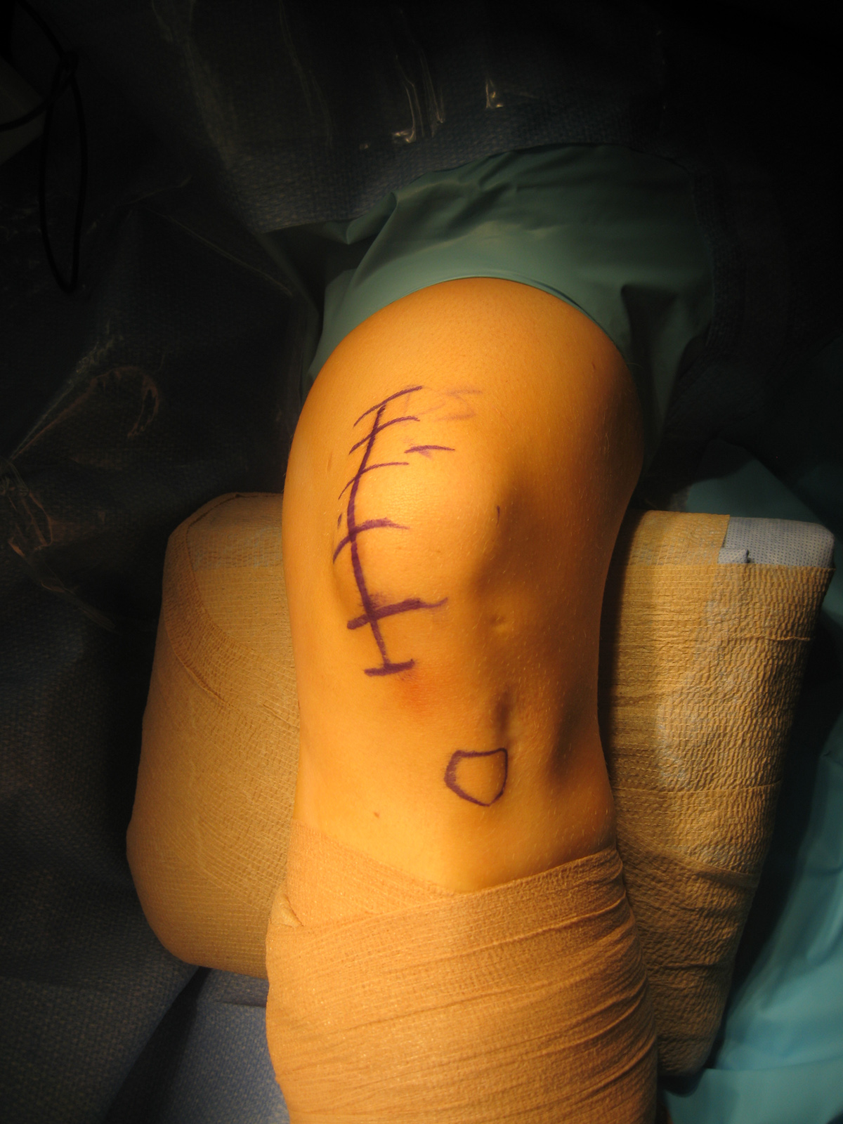
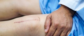
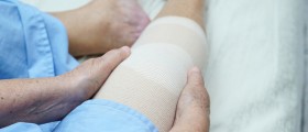
The surgeon first performs an arthroscopic examination of the joint. This is performed under general anesthesia and during the procedure makes necessary corrections. What follows is placement of a specific size of bone wedge in the upper part of the leg just above the knee. The bone is fixed with a plate and screws and the procedure lasts approximately 1 hour. There are no braces or plaster just bandage which is removed on the third day after the surgery. Patients may start moving their legs immediately. The drainage tubes stay in the wound for one day.
- Of the patients requiring a primary total knee arthroplasty (TKA), 10% to 15% present with valgus deformity (VD), the inaccurate correction of which continues to be a challenge for the orthopedics even currently.
- The valgus knee may have any combination of primary or secondary abnormalities even osseous (acquired or preexisting bony anatomic deficiencies) or soft-tissue (lateral and medial). These include on the one hand contraction of the lateral capsule and lateral soft tissues and ligaments; and on the other hand lax medial structures.
- To understand the important anatomic changes in valgus deformed knees is absolutely helpful so as to choose the best surgical method, to optimize correct component position and restore gap and soft-tissue balancing.
- After the clinical assessment, the mandatory pre-operative planning radiographs that should be included are: (1) a weight-bearing knee anteroposterior view; (2) a lateral; and (3) a patella merchant view.
- In the last three decades, a number of different surgical techniques have been described for TKA, in severe valgus deformed knees. As already mentioned, with the aim of correcting the mechanical axis in valgus knees and achieving soft tissue stability, proper bony alignment and ligament balancing are critical. The distal femoral cut at 3° only, instead of 5° to 7° that applies in varus knees, protects against under-correction. In order the mechanical axis after operation not to shift back into valgus, a slightly more varus result has been proposed during TKA for VD.
- Above and beyond, on the subject of ligament balancing in valgus knee there is no consensus on the subject of the correct sequence in which the lateral elements should be released.
- Very often, especially in cases of tibial tubercle osteotomy, there are operative or post-operative proximal tibial stress fractures. These fractures can be treated surgically or conservative including application of functional knee brace and toe-touch weight bearing of the affected leg till the fracture heals. Peroneal nerve palsy has been cited as an important complication after TKA for VD. The elongation of the lateral side stretches the nerve and places it at risk for indirect injury, via traction or induced ischemia.
After the surgery patients are explained specific exercises by physiotherapist. These exercises are a significant part of proper rehabilitation. In the beginning patients will use crutches for walking trying not to put much pressure onto the operated knee.
Possible complications of this surgical procedure include bleeding and infections. In case of surgery failure patients will undergo additional surgeries or bone grafts. The plate which is used to fix the bone is actually beneath the skin and it may irritate the overlying soft tissues. The plate may also restrict the range of motions. If there are problems like these the plate will need to be removed. The improvement after the operation is gradual and it lasts approximately a year.








Your thoughts on this
Loading...