Labrum Cartilage and Its Damage
The labrum is a cartilage located in the shoulder joint. The very joint contains two types of cartilage, the articular cartilage and the labrum. The labrum cartilage differs from the articular cartilage since it contains more fibrous tissue which makes it more rigid.
The labrum cartilage is normally found around the socket. This is actually the place of its attachment. The labrum cartilage is a very important cartilage. It has two vital functions. The first one is to deepen the socket and this way allows the ball end of the humerus to be in its place. Secondly, this cartilage represents a place where other structures and tissues of the shoulder joint attach.
The labrum can be torn and there are several places where this injury can occur. The tear can occur inside of the labrum (this is usually a small tear). Furthermore, the tear can develop at the place where the cartilage meets the biceps tendon. A complete tear is usually associated with a sub-luxation of the shoulder joint.
Labrum Surgery - Recovery Time
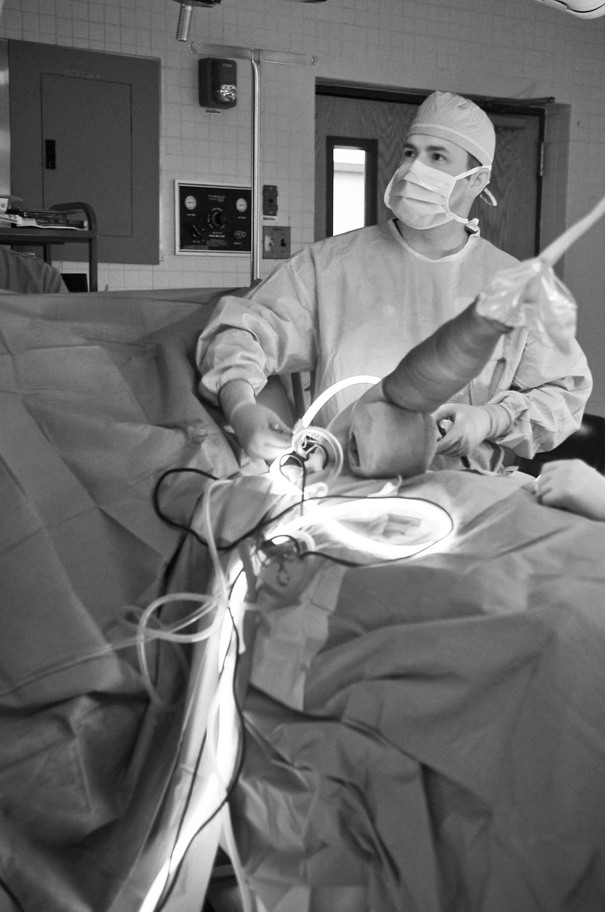
The treatment from labrum tear in majority of cases includes surgical repair. Surgeons most commonly opt for an arthroscopic surgery. This is a very successful surgical procedure.
The recovery time after labrum surgery is the time necessary for the shoulder to completely heal and regain its functions. Recovery time after labrum surgery basically depends on several factors which interfere in the actual speed of recovery. In general this lasts approximately 3 to 5 months.
After being discharged all the patients are due to follow doctor's orders and do not engage in any kind of strenuous activity that will put too much pressure onto the operated joint. The first visit to the doctor after the surgery is usually after a week. The surgeon checks on the incision line. This way he/she make sure that the wound has not been infected. This is also the most convenient time for a patient to engage in physical therapy. The patients are explained and taught how to perform specific motion exercises. These exercises are adapted to range of motions allowed after the surgery. Within first two weeks the arm may be placed in a sling. The person must not go too far with exercises and once the pain occurs it is a sign for a patient to stop. Light exercise should never be painful.
The doctor decides when the patients can start participating in more complex and strenuous physical activity and different sports. It is essential for all the tissues of the shoulder to heal properly and all the shoulder muscles to regain their strength prior engaging in any kind of more complex physical activity.
- Participants of a study completed a pain Visual Analogue Scale (VAS), the Veterans RAND 12-item Health Survey, Western Ontario Rotator Cuff Index, American Shoulder and Elbow Surgeons instrument, Scapular Assistance Test (SAT), Shoulder Activity Level, and Single Assessment Numeric Evaluation at baseline and at 6-month, 12-month and 2-year follow-ups. ?2and Student's t-test were used to test the differences between categorical and continuous variables. Analysis of variance investigated the differences between groups, and linear regression analyses explored the relationship of baseline characteristics with outcome scores.
- After 2 years, the operative cohort (n=68) significantly improved in all measures. The non-operative cohort (n=55) showed significant improvements in all scores except the mental component summary (MCS) and pain VAS. Labral tear patients (n=52) within the operative group (n=28) significantly improved in all measures except MCS. Non-operative labral tear patients (n=24) indicated significant improvements in all measures except MCS, VAS and SAT. Labral degeneration patients (n=71) within the operative group (n=27) significantly improved in all measures except MCS and SAT. Non-operative labral degeneration patients (n=44) indicated significant improvements in all measures except the physical component summary, MCS, VAS and SAT.
- In this patient population, pain VAS was significantly higher in the operative group at baseline. However, there was evidence to support the efficacy of surgical intervention as these patients electing operative treatment displayed significant improvements from baseline to 2-year follow-up across all outcome measures. While the non-operative group also reported significant improvements in outcome measures, the operative group exhibited larger MDs from baseline to follow-up, as well as higher scores associated with better outcomes. Additionally, the reported raw 2-year follow-up scores were significantly higher/improved when compared with the non-operative group except for MCS and SAT.
- VR-12 (MCS and PCS) scores were not significantly different at baseline between operative and non-operative groups. Despite observing an increased MCS score in both operative and non-operative groups, there was no statistical difference between 2-year MCS scores of either treatment group. Throughout this study, MCS consistently did not display significant improvements over time. This phenomenon is not fully understood, but can possibly be attributed to the nature of a repaired labrum.








Your thoughts on this
Loading...