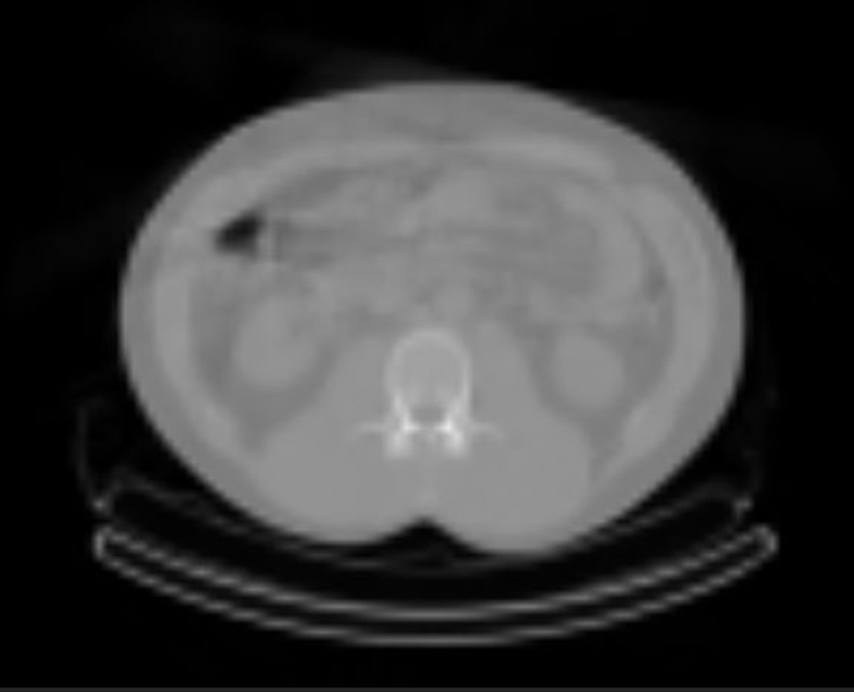
Chest CT scan
A chest CT scan is used for various reasons. However, some of the main reasons why doctors use them are to get the real size, shape and position of the lungs and every other structure located in the chest and to discover the real cause of lung symptoms like shortness of breath and chest pain, among others. If certain out of the ordinary findings were spotted on a standard chest x-ray test, a CT scan is ordered. Discovering if a person suffers from some of the many lung problems like a tumor, too much fluid around the lungs and a pulmonary embolism is also done through a chest CT scan. In situations when a doctor is suspecting of pneumonia, tuberculosis or emphysema, he can properly diagnose it after a CT scan.The machine that performs the scanning does not take only one picture of the lungs but a lot of them. These pictures, also known as slices, are then processed by the computer and the doctor is able to show them to the patient on a screen or a print them on film. A highly detailed 3D model of the organs can also be seen on the screen. The doctors often inject a certain substance into the vein of the patient before he or she goes for a CT scan. The name of the substance is contrast dye and it is given in order for a clearer image to be gained as the areas in the chest will be highlighted due to it.
Because there is need for more than one diagnostic use of a chest CT scan, there is more than one type of a chest CT scan. High-resolution CT scans or HRCT are a good option as a single rotation of the x-ray tube provides more than one picture of the chest. Every picture contains a lot of information and details about the chest and all the organs located in it. In case of spiral chest CT scan, a hole shaped like a tunnel is used as the x-ray tube rotates around the patient. At the same time when the tube rotates, the table moves through the hole. Thanks to this movement the x-ray beam is able to follow a spiral path. This type of a chest CT scan is used when there is need for a 3D image of the chest and the structures inside it.A chest CT scan and the preparation for it do not last for longer than half an hour. The most of the time is spent on the preparation while the actual scanning does not last longer than a couple of minutes. When undergoing a CT scan it is vital that a person remains calm and does not move as the picture can become blurry if he or she moves. In some situations a patient is even asked to hold the breath for a couple of seconds in order for the picture to be perfect. As some patients feel anxious and nervous inside closed spaces, the doctors often give certain medications that help them relax.
Are there any risks involved?It is not often seen that a CT scan leads to side effects. However, certain risks do exist. As a CT scan uses radiation, there is a slight chance of developing cancer but that almost never happens. The experts are still unsure just how much a CT scan increases the risk of developing cancer. The amount of radiation depends on the type of CT scan. The radiation is no longer present in the body after only a couple of days after the test is being performed. More precautions are needed when children are the ones than need to be scanned because they are a lot more sensitive to radiation than adults. The doctor will lay out all the possible risks and benefits of a CT scan before it is ordered.
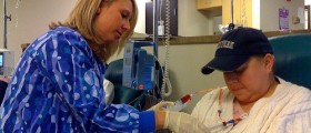
Exposure to radiation is not the only risk that can happen during a CT scan as an allergic reaction can occur due to the contrast dye that is being given before the scan. Hives and difficulty breathing occur most often. As most contrast dyes contain iodine, a patient needs to inform the doctor if he or she is allergic to it.








Your thoughts on this
Loading...