What is Bone Mineral Density?
The amount of mineral matter per centimeter of bone is known under the name bone density. The term is generally used in clinical medicine and is the major indicator of medical condition called osteoporosis and the accompanying problems such as susceptibility to fractures. Bone density shows how strong our bones actually are. This variable is evaluated with the assistance of densitometry, a procedure performed in nuclear or radiolography medical departments. The average value of bone mineral density is approximately 1500 kg m−3.
The procedure is painless and non-invasive but includes some exposure to radiation. This exposure, however, is not high. In the majority of cases measurements are performed on the lumbar portion of the spine or over the upper portion of the hip bone. If both of these body parts are not suitable for measurements the doctors scan the forearm.
According to data bone density reflects the probability of bone fracture. Namely, in individuals with low bone density bones are not strong enough and they are prone to fractures, particularly leg bones and the pelvis.
So, people who are assumed to have low bone density according to their medical record are due to undergo bone density scan which will either confirm the assumption or rule it out. Those confirmed with low density, osteopenia or osteoporosis are then prescribed specific treatment which may comprise dietary changes, calcium and vitamin D supplements along with weight-bearing exercises or even medications (e.g. calcitonin, Alendronate, ibandroonate etc.).
Importance of Bone Density Scans
Bone density scan is a powerful tool which efficiently evaluates the amount of bone in the particular part of the body. The test is generally performed on the spine, hip, forearm or rarely the heel. The goal of the procedure is to evaluate the probability of future fractures i.e. assess whether the bones of the tested individuals are 'weak and fragile' and can be easily broken.
The risk of fractures is basically increased in individuals suffering from osteoporosis. Their bones do not contain sufficient amount of calcium and other minerals and are not strong enough to withstand certain forces. The information obtained with bone density scans is sufficient enough for doctors to confirm/rule out osteoporosis and additionally determine the degree of bone weakness which certainly plays significant role in future treatment.
Apart from determining density of the measured areas, bone density scans also allow medical experts to predict future osteoporotic fractures at other areas of the body.
All in all, bone density scans are definitely priceless. This is a great way to diagnose osteoporosis on time and begin with treatment instantly.
Diagnosing Osteoporosis with Bone Density Scan
Osteoporosis is, by definition, a medical condition, to be more precise a bone disease characterized by a reduction in bone density and subsequent increased risk of fractures. One of the greatest problems associated with osteoporosis is that people are asymptomatic and only once they experience a fracture or repeated fractures and undergo additional tests they are confirmed with the disease.
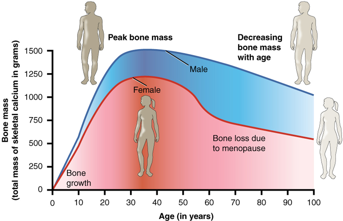
It is estimated that 44 million people living in the United States are suffering from osteoporosis and low bone density. Women are in general more susceptible to the condition especially when they enter menopause when the ovaries reduce the production of certain hormones essential for maintaining healthy and strong bones. Additionally, people at risk include those with a family history of osteoporosis, individuals of short and thin stature, people leading sedentary lifestyle and those prescribed some medications such as steroids.
Currently dual energy X-ray absorber is the most frequently used method of measuring one's bone density. The machine emits low radiation, There are two beans, the first one of high energy and the second of low energy. Specifically determined amount of X-rays passes through the exposed part of the body (including the bone tissue) and it is then measured. So, by comparing the difference between the two beams the doctors finally get the information about the thickness of the tested bone.
This test is generally performed on the spine and hip and can be highly predictive about potential future fractures. Scanning lasts no longer than 20 minutes, is completely painless and is associated with no complications.
Only patients who are able to lay still on the table undergo testing. There is no need for intravenous line because no contrast dye or any medications need to be administered. Patients may eat and drink practically everything on the day of the examination. The only thing they should avoid 24 hours before the test are calcium supplements.
The reliability of the test may be jeopardized by certain conditions such as scoliosis (a deformity of the spine), atherosclerosis and the presence of multiple fractures.
And finally, many people wonder how often the scans should be done. One scan is sufficient for most patients and it will precisely assess the overall risk of potential future fractures. A second scan can be performed after 2-3 years in case doctors want to make a new decision regarding changes of the current treatment.
Your thoughts on this
Loading...