
CT scanning, also known under the name CAT scanning is a diagnostic procedure that gives insight in specific body structures and tissues once these are exposed to X-rays of specific quality. The procedure is completely non-invasive. The only invasive thing related to CT scans is administration of a contrast dye which efficiently enhances the quality of the obtained images.
What is a CT Scan?
CT scans are of major importance for diagnosing a variety of medical conditions and they are also performed when doctors evaluate the efficacy of treatment or check for the recurrence of the disease.
The images during CT scanning are obtained with the assistance of the combination between sophisticated computer systems and special X-ray equipment. The final result of scanning are cross-sectional images of the inspected area and they are either displayed on a computer monitor or printed. They may be also stored on a CD.
The very procedure is precise enough to be more opted for than a regular X-ray which gives only 2-dimensional insight in the problematic area while CT scans may easily visualize the problematic region in all three dimensions allowing doctors to be more sure when diagnosing the underlying health issue. Also, the clarity of certain tissues, organs and blood vessels is much better compared to standard X-ray which sometimes cannot visualize the desirable structures.
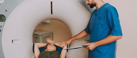
The CT scanner is actually a fairly large machine resembling a box with a hole in it. The hole is more like a short tunnel. In the middle of the hole/tunnel, there is a table which can be easily moved, carrying the patients to portions where the desirable body parts are exposed to X-rays. The X-ray tube rotates around the body while electronic X-ray detectors collect information which is later transferred to the computer workstation. The station is never located in the same room as the machine. It is in the room that is separated with a large glass window which allows medical staff to manipulate the machine and still have insight in what is going on during the exam. Also, monitors of the scanner soon after scanning show the images of the inspected parts of the body.
Head Injury Diagnosed with CT Scan
Traumatic head injury is a relatively common medical condition affecting people of all ages. It develops as a result of an external force which is blamed for traumatic injury to the brain as well as other structures in the cranium such as cranial nerves and various blood vessels. This type of injury is responsible for death of many people and is also blamed for permanent disability, particularly in children and young adults. In the majority of cases the injury is associated with car accidents, accidental falls and violent behavior. Even though technology has progressed significantly over the years, it is still not possible to to fully protect people who drive cars, motorcycles or participate in sports that carry increased risk of traumatic brain injuries.
The trauma the brain experiences develops due to direct impact or may be closely connected with acceleration. Apart from the direct damage, there are additional health issues which tend to develop over the course of several minutes or days after the injury. Also, secondary edema of the already damaged brain tissue increases the severity of the existing health problems and is many times blamed for lethal outcome.
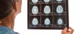
Fortunately, apart from physical and neurological exam, patients who have had traumatic head injury undergo CT scan of the head which gives insight in the site of damage, the extent of damage and its correlation with patient's symptoms and signs. For instance, CT scan of the head may easily reveal intracranial bleeding, subarachnoid hematomas, skull fractures, blood clots, compression of the cranial nerves etc. What is more, it is a rather powerful tool for evaluation of all the damage caused by facial trauma. It allows doctors and surgeon to plan further treatment and choose between conservative approach or perhaps surgery. CT scan also helps doctors determine which patients require immediate surgery.
CT scan is the first imaging technique doctor opt for when head trauma is concerned. The exam is quick and rather accurate. Still, in case CT does not provide with sufficient information, patients may need to undergo MRI. However, MRI is not used in emergency situations. It is also not as good as CT scan when it comes to visualization of bleeds and fractures, common injuries associated with head trauma.
It is of utmost importance to remove all the metal objects a casualty is wearing such as jewelry, dentures etc. because these easily affect the CT images and may prevent doctors from noticing the underlying injury.
All in all, traumatic brain injury is a serious medical issue which may cause permanent disability and a variety of neurological sequelae. However, thanks to imaging methods such as CT, predominantly, and MRI the injury of this type can be properly evaluated and patients treated within optimal period. Timely treatment may prevent many complications and help patients overcome the injury, achieving full recovery.




-Test-And-What-Do-The-Results-Mean_f_280x120.jpg)



Your thoughts on this
Loading...