Introduction
Nose bleeding may be caused by many different factors. However, most of these have one thing in common. Most of the nosebleeds are caused by bleeding from either an artery or a vein located somewhere in our face, our nasal tract, eye sockets or perhaps inside our head. If the bleeding is strong and cannot be stopped, there is a high possibility that it is caused by a bleeding artery, since, in these, blood pressure is much higher than in the veins.
First Aid and Surgery
In most cases, the bleeding can be stopped by the burning, freezing or applying electrical treatment to the troublesome spot. Nevertheless, in some, more complex cases, surgery is needed where doctors must separate the bleeding blood vessel from the nasal area or just simply block it. Under both of these circumstances, anesthetics are given to the patient since those are able to slow down the bleeding and remove any sensations of pain.
Sometimes, arteries need to be tied off, since only one of many may be causing the problem. This is done by operating the area spreading from our nose to our eye sockets. It is a very complex procedure and there are even cases when it is performed in the neck area as well as the cheek area.
- In an attempt to avoid complications during surgery, angiographic embolization for treating posterior epistaxis has first been described in 1974. The success of this procedure has been classically reported to be 71–95%. In a recent study comprising 70 patients who underwent angiographic embolization of the sphenopalatine artery, 13% had recurring bleeds within 6 weeks of the procedure and another 14% at a later presentation.
- The complications of this procedure have been extensively reported in the literature and include hemiplegia, ophthalmoplegia, facial paralysis/paresthesia, blindness, or other neurological deficits caused by accidental embolization of cerebral arteries. These possible complications were shown to occur in up to 27% of cases.
- In 1965, Chandler and Serrins described the transantral ligation of the maxillary artery under local anesthesia. This technique is classically performed through the Caldwell-Luc approach. It has been associated with persistent pain in the upper teeth, infraorbital neuralgia, oroantral fistula, sinusitis, potential damage to sphenopalatine ganglion and vidian nerve, and, rarely, blindness. Complications of this approach have been estimated to reach 28%
- A study conducted in 2006 suggested the use of endoscopic anterior ethmoid artery ligation only when the artery is in a mesentery and is clearly visible (present in 20% of cases according to the study). Otherwise, the authors rather suggested an external approach.
- Cautery under endoscopic vision is another option for the control of epistaxis that may avoid the uncomfortable insertion of nasal packs in the case of an unidentified bleeder. While this can be performed in the operating room, a well-equipped clinic or emergency department can also be adequate settings for this procedure. While some authors report a very high success rate, others report a relatively significant risk of failure (17–33%), which may be due to the fact that cauterizing the nasal mucosa may also damage an area that will bleed persistently.
- Endoscopic Ligation of the Sphenopalatine Artery or the ESPAL was first described over 20 years ago. Interrupting the blood flow in a sufficiently distal area provides an advantage over the previously described techniques by avoiding the possible revascularization from the internal maxillary artery.
- The management of posterior nasal bleeds will depend on the availability of experienced endoscopists and relevant equipment. The experienced endoscopist may be successful in treating these patients in an emergency setting, therefore avoiding the potential adverse effects of packs insertion and the potential complications and costs of surgery under general anesthesia. ESPAL can always be done after failure of this procedure.
Embolization of the Arteries
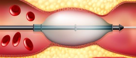
This stands for another, quite complex procedure. Namely, this is performed in the X-ray department and in most cases a radiologist does it. A needle is placed in an leg artery along with a special tube. This needle is then pushed forward all the way to the nose, releasing special dye which is visible under the X-ray scanner. This dye helps the doctor with finding the rupture or the bleeding spot in the artery. After the spot has been found, an adequate surgical procedure is conducted in order to isolate or stop the bleeding part of the artery.
Characteristics of the Procedures
Depending on the type of surgery, it may last from 10 or 20 minutes for the simple ones all the way up to 90 minutes and more for the complex ones like the above mentioned embolization. If there are no further problems or reoccurring bleeding issues, the patient is released after approximately two days.
Post-operative precautions may involve one's restraining from nose blowing or lifting heavy things as well as the necessity of using nasal sprays or some similar medication prescribed.
Unwanted side-effects involve numbness due to nerve damage, danger of blindness, bruising of certain spots or some allergic reactions. However, in most cases the operations tend to be fully successful.


Your thoughts on this
Loading...