Eye lesions represent a loss of integrity of certain eye structures and their layers. They can be sometimes fatal and result in loss of vision. Even though it is estimated that approximately 80-85% of eye lesions are actually benign, the rest of them can be associated with serious medical problems.
This is the reason why any new lesion on the eye, any change in color or shape must be reported and one should be thoroughly examined.

Why do Eye Lesions Occur?
There are many reasons behind eye lesions. Most of them can be easily brought under control with antibiotics or some other eye drops or creams, warm compresses, and surgery.
Certain lesions are closely associated with irritation, while others may be induced by exposure to harmful UVA and UVB sun rays. It is also possible to end up with an eye lesion after exposure to some pathogen. All in all, waiting for too long and letting the lesion spread and enlarge is not a wise thing to do, so do visit your doctor as soon as you notice anything strange about your eyes.
Frequent Eye Lesions
A nevus is a benign eye lesion that can also be found anywhere else in the body. If affecting the eye nevus can or does not have to be pigmented. This is a congenital lesion often considered harmless. However, if a nevus changes its color, starts to grow, etc. this is a reason for one to consult his/her doctor.
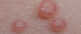
Many viruses can cause inflammation of the eye and eye lesions. Herpes simplex virus type 1, for instance, predominantly affects the cornea and leads to the formation of superficial or deeper corneal lesions (ulcers). Furthermore, there is a condition known as Molluscum Contagiosum. It basically affects immunocompromised individuals and develops in a form of a waxy nodule. The lesion will not withdraw on its own and is removed with cryotherapy.
Actinic keratosis is a pre-cancerous lesion affecting different parts of the body and also develops on the eyelids. This growth resembles a horn and is flaky and basically white. It develops as a result of too much exposure to UV light. Actinic keratosis requires surgical excision because if left untreated, it eventually progresses into squamous cell carcinoma.
Seborrheic keratosis is a benign lesion affecting the eyelid. It does not disturb the affected individual and can be, if necessary, surgically removed.
- Persons ranging in age from 43 to 84 years and living in Beaver Dam, Wisconsin, at the time of a census (1987–1988) were examined two to three times over a 10-year period (n = 3684). Drusen area, size, and type; retinal pigment epithelium depigmentation; increased pigment; geographic atrophy; and neovascular macular degeneration were determined in each of nine macular subfields: central, inner and outer superior, inner and outer nasal, inner and outer inferior, and inner and outer temporal by grading of stereoscopic color fundus photographs. Late ARM was defined as presence of either geographic atrophy or neovascular ARM.
- Lesions were more likely to change or develop in specific locations. Drusen area increased most in the central circle. Compared with other quadrants, drusen greater than 125 ?m in diameter and soft indistinct or reticular drusen were most likely to develop in the superior or temporal quadrants, whereas pigmentary abnormalities were most likely to occur in the nasal or superior quadrants.
- In general, large drusen, soft indistinct drusen, and pigmentary abnormalities were more likely to develop in the inner circle versus the central and outer circles. The quadrant location of early ARM lesions in 72 persons in whom late ARM developed was generally similar to that in persons who did not have late ARM.
- However, persons who had geographic atrophy were more likely to have large drusen in the inner circle than in the outer circle, while those who did not have late ARM were more likely to have large drusen in the outer circle.
Hydrocystoma is a lesion associated with a blockage of sweat glands. It is also removed surgically (excision).
Malignant lesions, on the other hand, require a more complex approach and treatment. The eye area may be affected by three malignant tumors, squamous cell carcinoma, basal cell carcinoma, and melanoma.
Squamous cell carcinoma is rather aggressive and develops in a form of raised, the scaly lesion usually on the upper lid. Basal cell carcinoma is also a malignant tumor, but it progresses very slowly. Surgical treatment deals with the tumor and provides complete eradication of the disease. Malignant melanoma is one of the most aggressive tumors and even in the early stage, it can give satellite metastases in the surrounding tissue.
Finally, the eyelids may be affected by one more malignant tumor called sebaceous carcinoma. This is an aggressive tumor that, if left untreated, may easily spread to distant organs such as the lungs, liver, and bones.










Your thoughts on this
Loading...