
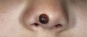
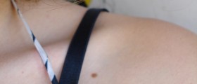
A melanocytic nevus is a skin lesion originating from melanocytes (melanin-producing cells found in the skin's epidermis, the middle layer of the eye, the inner ear, meninges, bones and heart). A melanocytic nevus is of brown or black color due to high concentration of melanin, a pigment synthesized by melanocytes. It is confirmed that the majority of moles develop and occur during the first two decades of a person's life and that certain number of babies (approximately 1 out of 100) is born with these skin lesions. Unlike acquired nevi, which are considered benign neoplasms, congenital nevi represent a risk factor for melanoma.
Melanocytic Nevi Classification
There are several variants of melanocytic nevi.
A junctional nevus is generally flat, brown or black and forms along the junction of the epithelium and the underlying dermis, hence its name. A compound nevus is a form of junctional nevus with intradermal proliferation. Such nevus is slightly elevated and also brown or black. Intradermal nevi are located in the dermis, are always raised and in majority of cases have no pigment.
Dysplastic nevi are characterized by dysplasia, they are either flat or raised and basically vary in size. In many cases such nevi are large and must be removed because they may eventually transform into skin cancer.
A blue nevus comprises melanocytes buried deep in the skin. Such nevi are spindle shaped. Spitz nevi occur in children, are raised and red.
Giant pigmented nevus, as the name suggests, is a large and most commonly hairy congenital skin lesion. These nevi are also potential precursors of melanoma.
Intramucosal nevi affect mucous membranes of the oral cavity and the genital area.
Nevus of Ito as well as nevus of Ota are congenital nevi. These skin lesions are flat and brown and basically form on the shoulders or face.
Mongolian spot is another type of nevus affecting Asian babies. This is a deep, congenital blue nevus.
Finally, there are recurrent nevi. They occur after incomplete removal of some nevus. The remnant melanocytes start to multiply and form a new nevus.
The Occurrence of Nevi
It is estimated that nevi appear during childhood and may gradually disappear after middle age. Congenital nevi, however, do not disappear. People of fair complexion generally have around 30 moles. The number can be much higher reaching up to 400 moles.
It is believed that genes can have an influence on nevi formation. For instance, dysplastic nevi as well as atypical nevi are hereditary conditions. In such individuals there are 100 or even more nevi some of which are quite large. These two conditions are closely associated with a higher risk for melanoma. UV radiation is apart from premature aging also connected with the formation of acquired moles. Excessive and unprotected exposure to sunlight is also a huge risk factor for melanoma.
All in all, whether nevi are congenital or acquired, any change in their appearance must be reported as soon as possible. A well-experienced dermatologist will exam all the nevi one has and will suggest removal of some of them (if necessary). This way one may prevent the onset or even progression of skin cancer.









Your thoughts on this
Loading...