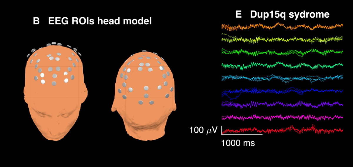
The human brain is a highly complex organ. Many of its functions are still a mystery to medical experts and yet to be explained. As for medical conditions affecting the brain, these can be confirmed in many ways. A family history and a neurological examination will provide with plenty of information. Structural changes are confirmed and evaluated with CT scan or MRI while EEG (electroencephalography) provides with information regarding the electrical activity of the brain.
Electroencephalography for Electrical Activity
Electroencephalography (EEG) is a relatively simple, non-invasive test which measures electrical activity of the brain with the assistance of electrodes placed on the scalp. The test has huge diagnostic value especially when various neurologic disorders are concerned. It is also performed after some brain surgeries and to check whether the administered treatment is successful.
EEG provides with plenty of information and is most definitely the tool of great power for both research and diagnosis, particularly when there are no obvious changes confirmed with CT or MRI. The test has its basic form and also some derivations. One of these is evoked potential, a specif type of EEG where the results of the test reflect the response of the brain to various stimuli e.g. visual, Somatosensory or auditory stimuli.
As for clinical use EEG is a routine test which lasts no longer than 30 minutes (including the preparation) and requires no anesthesia. Such, routine EEG is performed when doctors want to differentiate epileptic seizures from psychogenic non-epileptic seizures or other spells like syncope and migraines. Furthermore, EEG may distinguish ‘organic’ encephalopathy from primary psychiatric syndromes. The test also confirms brain death, coma and is of great significance when the success of current treatment is evaluated.
Additional indications for EEG are evaluation of the depth of anesthesia, effects of sedatives/ anesthesia in patients with medically induced coma and secondary brain damage due to subarachnoid hemorrhage and similar damage to the brain.
Is Electroencephalography Safe?
This is a highly safe test. The electrodes are placed on the surface of the scalp and are never inserted into the skin. There is no need for anesthesia since the procedure is neither painful nor it causes any discomfort.
However, there is a specific preparation for the test patients should follow. First of all, they must report all the drugs they are currently taking. Some medications may change the brain activity and interfere with the results. This is why once the doctor gets fully informed about the drugs the one is taking on a regular basis he/she will decide which of these must be discontinued prior to the test. Sedatives, muscle relaxants, antiepileptic etc. are all drugs which must be discontinued prior to EEG.
In addition, foods and drinks containing caffeine should not be introduced into the body for at least 12 hours before the test.
Also, clean hair, without any presence of sprays, oils, creams and similar is a must since electrodes have to stick to the scalp and the aforementioned substances may interfere with the connection between the electrodes and the surface of the head.
Since sometimes EEG must be performed when patients are asleep, these individuals are asked not to sleep the night before the exam or at least to sleep less than usual. This ways they will be tired and easily fall asleep during the exam. Such patients also should have someone who will drive them to the test and home because sleep deprivation in general makes one incapable of focusing and driving correctly.
All hospitals and many doctors’ offices are equipped with EEG machine. The test is performed by a well experienced EEG technologist and evaluated by a neurologist, a medical expert specialized in the diagnosis and treatment of neurological disorders.
Patients either lie on their back or sit in the chair. There are 17-21 electrodes attached to specific parts of the scalp. They are attached to the skin with a sticky paste. In some cases the electrodes are a part of the cap which is placed on the head and attached to the scalp with tiny needles.
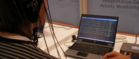
Electrical impulses generated by the brain are received by electrodes and then sent via wires to a computer. The result is in the form of a series of wavy lines which are displayed on the computer screen or may be additionally drawn by a row of pens on a moving paper.
Naturally, patients are supposed to follow the technologist’s orders. For example, they should not to speak unless they are told to. Also, some of them must fall asleep for the purpose of thorough examination. There are many other activities performed during the testing which contribute to the diagnosis of the underlying condition.
After the test doctors evaluate the results and according to changes on EEG opt for the most suitable treatment. Patients may go home immediately or wait for their doctor’s report. All in all, there are no limitations after the exam except for EEG performed after sleep deprivation when patients should have someone to drive them home.









Your thoughts on this
Loading...