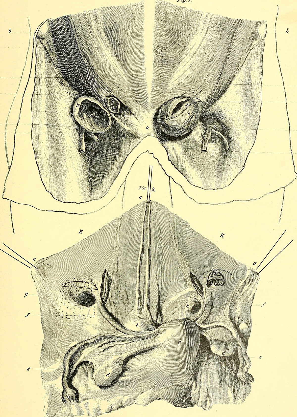
A femoral hernia is a condition that occurs when feebleness of the groin muscles allows for the intestine to bulge through. An unorthodox bulge in the groin or upper thigh area is usually a good sign of this condition.
Discerning a femoral hernia from an inguinal one may prove to be a tough task. The only difference between the two is their position relative to the inguinal ligament. A hernia located above the inguinal ligament within the groin area is an inguinal type. Below the inguinal ligament would be the femoral hernia. It is very important that an exact diagnosis is made only when the surgery begins.
A femoral hernia can be mild enough to only push the peritoneum or the lining of the abdominal cavity, through the muscle wall. More severe cases includes whole portions of intestines pushing through it.
This condition is caused by a number of things. Continuous, bowel movement inducing strains can cause it. The same goes for urinating strains, often resulting from prostate problems. A chronic cough may elevate the risk of developing this condition. Obesity is another possible cause.
Femoral hernia is most commonly found in women, though it is far from being exclusive to them. Older women and very small women are most often presenting within it.
This ailment will not go away without proper treatment. It requires surgery. The hernia may be small in the beginning, but it can grow, substantially, over time. Different activities may induce growth as well as shrinkage.
An incarcerated hernia occurs when the bulging content gets stuck. Although it is not considered to be an emergency, it should be addressed without unnecessary delays. A strangulated hernia, on the other hand, IS an emergency. It happens when the tissue gets cut off from the blood supply due to the narrowing of the hole in the muscle wall. Death of tissue is a likely consequence of this complication, making it very dangerous.
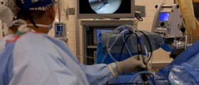
Surgery can be done on inpatient or outpatient basis. After giving the anesthesia, an incision is made on either side of the affected area. The pouch content is isolated and returned into its proper position. After this is done, a procedure to repair the muscle defect ensues.
Oftentimes, the defect may be small enough to treat it with suturing. This helps preventing the hernia from reappearing. A larger defect may require a mesh graft implementation. This is also a permanent solution as the mesh covers the hole completely, preventing re-occurrences. Using suture to cover larger defects elevates the risk of re-occurrence. Still, this may be necessary in patients who have a history of rejecting surgical implants of this kind.
After positioning the mesh, or sewing up the muscle, the laparoscope is removed and the incision is closed.
Recovery often takes no more than four weeks. The treated area will be tender initially. The incision should be kept protected during straining activities, through the application of gentle pressure. Activities that require this type of measure are sneezing, coughing, strenuous bowel movements and vomiting.





Your thoughts on this
Loading...