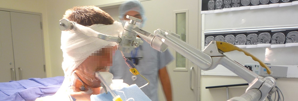
Craniotomy is a surgical procedure that includes opening the scull or cranium. After craniotomy, a surgeon has access to the brain and blood vessels in the skull, and then additional procedures such as tumor removal or operation of aneurysms can be done. Patients are generally administered general anesthesia. Still, they can be also awake and given local anesthesia in some cases. After the surgery, the hole in the skull is closed with a titanium plate or some other type of fixation.
After being operated the patient is monitored in the recovery room for a certain period. Vital signs are checked, and he/ she is then transferred to the intensive care unit. Nausea and headache are rather common after the surgery. The patients are administered special medications to reduce brain edema and prevent seizures. Hospitalization lasts from three days up to two weeks.
Craniotomy Complications
As with any other surgical procedure, craniotomy carries a certain risk of developing postoperative complications. Headaches are regularly present, therefore they are not considered a complication of the surgery. They tend to linger up to several weeks after the surgery. Seizures can develop as a consequence of cutting inside the brain. Still, one is prescribed anticonvulsants to prevent the seizure from occurring.
Before being discharged, a patient is given a list of what he/ she must do and what needs to be avoided by following the given rules majority of complications can be evaded.
In case of unexpected symptoms and signs, one should go to the doctor immediately. High body temperature is one complication. If it exceeds 101 Fahrenheit, patient must report it to the doctor.
Additionally, a patient has to report any case of redness, edema, or even pain in the operated area. If one notices sleepiness, problems with stability, or even skin changes, he/ she has to report these to the doctor. The last three symptoms may be connected to prescribed anticonvulsants.
Severe headaches, nausea, and prolonged or unstoppable sleepiness may point to increased intracranial pressure, which is an urgent state, and severe complication. Increased intracranial pressure may be induced by brain hemorrhage or the formation of blood clots. Reactions to anesthesia are not so common. Leakage of cerebrospinal fluid can be surgically repaired.
Unlike previously mentioned complications, which can be solved, some complications may lead to permanent damage of the brain tissue. They include stroke, seizures for a lifetime, damage to the nerves that can lead to irreversible paralysis of certain intracranial nerves, and permanent loss of mental functions.
- One of the most traditional craniotomies utilized is the pterional craniotomy, a supratentorial craniotomy that can be utilized for aneurysms of the anterior circulation, basilar tip artery aneurysms, direct surgical approaches to the cavernous sinus, frontal and temporal lobe tumors, as well as suprasellar tumors such as pituitary adenomas and craniopharyngiomas.
- The frontal craniotomy is used to access the frontal skull base and the frontal lobe of the brain for approaches to the third ventricle or sellar region tumors, craniopharyngiomas, planum sphenoidale meningiomas, frontal lobe tumors, and repair of anterior cerebrospinal fluid fistulas. Other types of craniotomies include parietal, occipital, and retrosigmoid, among many others.
- There are very few contraindications to performing a craniotomy: high anesthetic risks (advanced age, severe medical comorbidities), morribond state, poor functional status, high frailty index, severe systemic collapse (sepsis, multiorgan failure), coagulation disorders, bleeding dyscrasias, and pathologies that can be addressed by a single burr hole.
- Preoperatively, the patient must be in the best optimal condition possible to tolerate the procedure. The patient must be with an empty stomach or "nil per os" (NPO), a Latin phrase that translates to "nothing through the mouth" in the English language. In emergency cases, this is usually not possible. Blood-thinning medications such as antiplatelet or antithrombotics should be discontinued between 3 to 10 days preoperatively, depending on the drug.
- The reported incidence of major complications is 8.3%, with minor complications rising up to 60%. Mortality owing to major complications is reportedly 22% (0.5% in minor complications).
- Depending on the specific type of pathology to be addressed and the clinical judgment of the physician, it is determined if a craniotomy is necessary for the patient. Even with the new endovascular techniques to treat intracranial vascular disorders and the radiosurgery technique to treat intracranial tumors, craniotomy remains the primary tool for the treatment of the majority of neurosurgical pathologies.








Your thoughts on this
Loading...