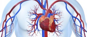
A thallium scan is a diagnostic test that is used to determine any problems that may be affecting the heart. It produces images of the heart muscle and it is usually combined with an exercise test. Its main goal is to determine if there are the certain areas of the heart that are not getting enough blood. Thallium scan is very useful in coronary artery disease.
Safety and the results of thallium scan
Thallium scan uses a radioactive substance called traces, which is injected into the blood stream and followed through the scan. Since the substance is radioactive, people can be less than willing to take this particular test, however the amount of radiation is very low and it all gets expelled by the kidneys in a short while. Thallium scan is therefore considered to be safe for most people, however, pregnant women should consult their doctor before undergoing this test.
When the tracer is injected into the bloodstream, the patient is asked to walk on a treadmill. The traces travels the bloodstream, through the coronary arteries and it is picked up by the heart muscle cells. It is picked up almost instantly in areas with normal blood supply. Areas that remain lighter and called “defects” are those that do not receive adequate blood supply and should be examined and monitored.
After the treadmill, the patient is asked to wait and to rest, during which time another set of images is taken. They determine which areas are temporarily defected, and which have suffered a permanent damage due to a heart attack.
During the test
A thallium scan is performed in a medical facility, a clinic or a hospital, usually at nuclear medicine or radiology department. It consists of two portions- exercise portion and imaging portion.
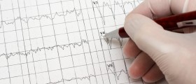
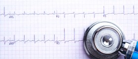
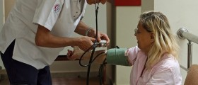
In the exercise portion, the patient is asked to step on a treadmill that at first goes slowly, but then gradually increases in speed and incline. During walking, the patient has electrodes that connect to an ECG machine, and cuffs that measure the blood pressure. The machine watches for abnormalities and the blood pressure is measured every few minutes.
The test stops when the patient is too tired to continue, when significant symptoms occur or when the heart rate reaches its peak.
After this, the patient is asked to be still and to rest. He or she will lie on a bed with a special camera above, which takes pictures at different angles. This usually takes 15 to 20 minutes.
The next scan is performed after two to four hours, during which time the patient can leave the area but should refrain from physical strain and from food as well. new pictures are taken, and they will show the heart in the resting state.
Doctors can give preliminary results to the patient while he or she is still there, but complete and accurate results will be ready in the following days.


Your thoughts on this
Loading...