Pupils
Pupils are holes in the iris of the eye. They allow the light to enter the eye and reach the retina. Normally pupils dilate if there is not enough light, and in bright light, they shrink. This occurs simultaneously and both pupils are involved in dilatation or constriction.
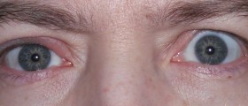
They are commonly even in size, and in some medical conditions, one can be dilated or shrink while the other stays normal. If the pupils are uneven in size, this medical condition is called anisocoria.
Symptoms of Uneven Pupils
Anisocoria may be mild, and sometimes the difference in size between the pupils can be drastic. The only sign is the uneven size of the pupils.

The cause is related to the muscles which are in charge of dilatation and shrinkage of the pupils. These muscles are innervated by the autonomous nervous system. Dilatation and shrinkage of the pupils are unconscious and cannot be controlled.
Some people suffering from uneven pupils may experience fear or pain of light, which is called photophobia. The eyes may be also exposed to increased strain. This leads to pain. Additional symptoms may include headaches and migraines.
Causes of Uneven Pupils
Uneven size of the pupils may be hereditary or acquired. There are numerous causes of this medical condition.
Certain eye drops, such as those used in allergies, may cause dilatation of pupils. Luckily eye drops only cause transitory problems, and there is no reason for the patient to worry about the unevenness of the pupils.
Certain plants may induce anisocoria if they come in contact with the eyes. These plants irritate the eyes, and rubbing the eyes can only make the situation worse. Some of them are Angel Trumpet, Deadly Nightshade, Yellow Jessamine flower, and so on.
Anisocoria commonly occurs in severe head trauma. It points to serious damage to the brain and its structures and requires prompt medical help and treatment.
In Horner's syndrome typical triad occurs. It includes ptosis, miosis, and enophthalmos. One pupil is smaller than the other. Horner's syndrome may occur as a consequence of damaged oculosympathetic nerve, sympathetic nerves that run down the carotid artery, etc.
In case the oculomotor nerve is damaged, the patient will have uneven pupils. Additional symptoms include double vision and drooping of the eyelids.
In Adie's Pupil, the actual cause of anisocoria cannot be identified. Some believe that viral or bacterial infection is responsible for the unevenness of the pupils. Still, in patients who are suffering from this condition, pupils sometimes spontaneously restore their normal size.
Additional causes of anisocoria include meningitis, encephalitis, brain tumors, and sometimes chest tumors. Apart from that, even abuse of some drugs, such as cocaine or marijuana, may cause anisocoria.
- Important etiologies of anisocoria include third nerve palsy, Adie pupil, pharmacologic mydriasis, pharmacologic miosis, traumatic mydriasis, physiologic anisocoria, and Horner syndrome.
- A dilated pupil can be tested pharmacologically. The muscarinic agent pilocarpine, both dilute (0.05-0.15%) and non-dilute (1 to 2%), acts on the neuromuscular junction of the pupillary constrictor to cause miosis. Dilute pilocarpine will cause constriction in a dilated pupil of greater than two weeks due to denervation of the neuromuscular junction. This previously was thought to help differentiate this form of mydriasis from TNP, but newer results cast some questions on this. If non-dilute pilocarpine fails to constrict the pupil, then the pupil is pharmacologically dilated.
- The prevalence of physiologic anisocoria is generally considered to be around 10 to 20%, which does not seem to differ greatly around the world. Physiologic anisocoria does not seem to have a sex predilection nor occurs at a specific age. Physiologic anisocoria is probably the most common cause. The prevalence of other causes of anisocoria is associated with the prevalence of the underlying condition.
- Distinct pathways control miosis and mydriasis (dilation of the pupil). The parasympathetic pathway causes miosis by activating the iris sphincter. These pathways arise within the brain stem and then extend along cranial nerve III to finally innervate the iris sphincter. This pathway is activated by the pupillary light reflex and accommodation.
- If a third nerve palsy is causing anisocoria, imaging is recommended to rule out a compressive lesion, especially an aneurysm, which can be acutely fatal. An aneurysm can be most effectively imaged with a computed tomography angiogram (CTA) or a magnetic resonance angiogram (MRA) of the head. If Horner syndrome is causing the anisocoria and a carotid artery dissection or aneurysm could be the cause, imaging is recommended. A CTA or MRA of the head and neck should be performed.
- The treatment of anisocoria depends on the underlying condition causing the condition. Most causes of anisocoria only require observation. A referral to a neuro-ophthalmologist, ophthalmologist, or neurologist may be warranted in cases that do not resolve. Acute onset anisocoria that is concerning for a compressive third nerve palsy or horner syndrome should be sent to the emergency department immediately for imaging.
- Anisocoria itself is unlikely to cause significant complications, although some do exist. A larger pupil may cause light sensitivity and visual aberrations. A smaller pupil may cause worsened visualization through a cataract, difficulty viewing the fundus during the posterior exam, or difficulty in cataract surgery. The main complication of anisocoria is not the difference in pupil size but the complications of the underlying condition itself.
- medlineplus.gov/ency/article/003314.htm
- medlineplus.gov/eyemovementdisorders.html
- Photo courtesy of Sophie Riches by Wikimedia Commons: commons.wikimedia.org/wiki/File:Dilated_Pupils.jpg






Your thoughts on this
Loading...