
Acoustic Neuroma
Acoustic neuroma is a benign tumor which originates from the vestibulocochlear nerve. It is actually made of so called Schwann cells that are basic components of myelin, the sheet that covers nerves. These tumors predominantly appear in the labyrinth, inside the brain and inside the brainstem. Acoustic neuromas affect adults and are rarely found in children.
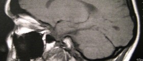
The tumor most commonly causes unilateral hearing loss and vertigo. It can be easily diagnosed by audiometry and MRI can provide with perfect insight of its localization and relation to surrounding structures.
In 50% of all the patients these tumors are surgically removed. In 25% of patients radiotherapy is applied. Some patients whose acoustic neuromas are small and do not cause significant trouble are monitored. Only if the tumor starts to grow rapidly or cause unbearable problems a patient undergoes the surgery or radiotherapy. The complication of both treatment modalities is significant of total hearing loss.
Causes of Acoustic Neuroma
Acoustic neroma may be a sporadic tumor. Apart from that in neurofibromatosis type II, which is inherited disease, acoustic neuromas may be only one characteristic of the disease. Luckily, 95% of all acoustic neromas are sporadic.
Surgery for Acoustic Neuroma
Surgical removal of the tumor has been used for many years. Still, this therapeutic method my be completely replaced with a new treatment modality - gamma knife. Open surgery definitely provides with better insight of the tumor and its behavior towards surrounding structures. This will help the surgeon to preserve the hearing as much as possible. Two specialists are required during the surgical procedure. They are neurosurgeon and neurotologist. The tumor can be removed by several surgical approaches.
The removal via labyrinth cannot help in preservation of hearing, but it also does not involve the opening of the cranium. This approach is rather convenient.
The second approach includes opening of the middle cranial fossa. This surgery may provide with preservation of hearing. On the other hand, since middle fossa approach includes opening of the cranium and manipulation with the brain and brain structures this approach carries a lot of risk for later complications.
Every manipulation inside the skull carries potential risk of damaging some of the brain structures. This is why a team of surgeons must be included in the process. The best option is total removal of the tumor. In some patients due to localization the tumor is only partially resected. Follow up includes regular examinations and occasional MRI of the cranium. This way the recurrence will be found on time and the patient may be re-operated.







Your thoughts on this
Loading...