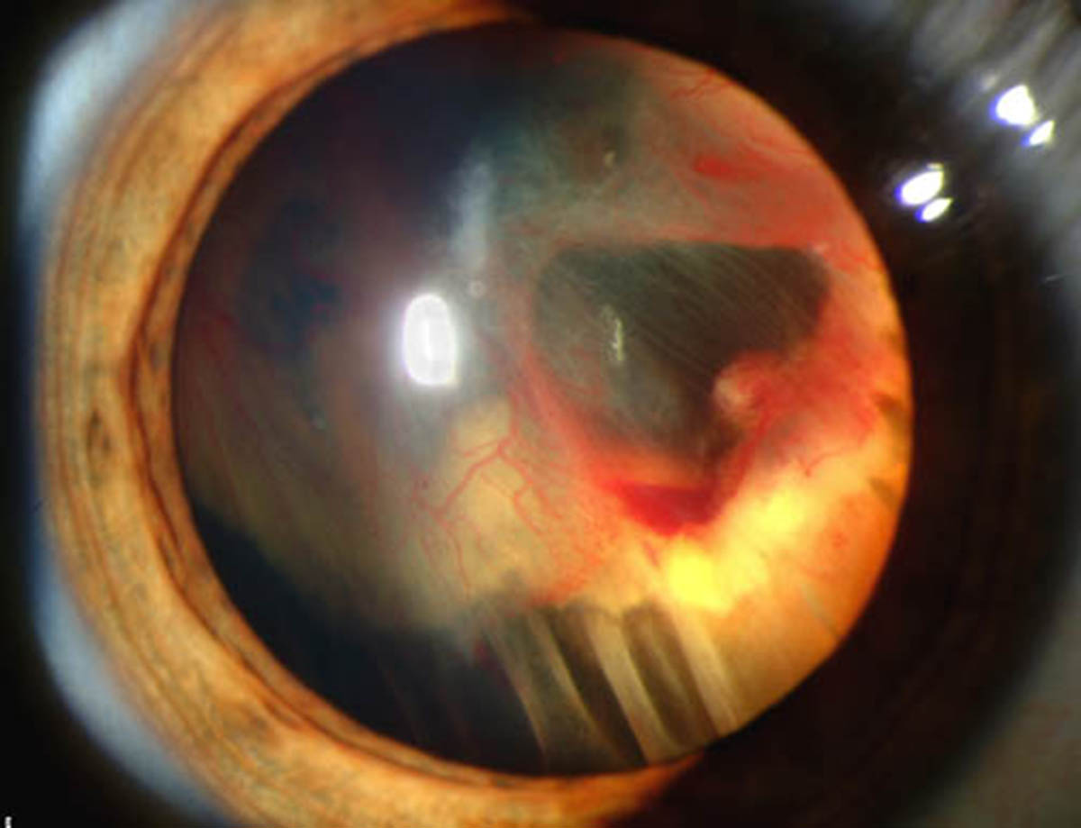
Retinal detachment is an eye condition affecting the retina, a light sensitive tissue of the eye in charge of light reception and further transmission of light signals to the brain. In case of detachment the retina separates from the back of the eye and leaves its supporting tissues. The condition is severe and must be treated promptly. Left untreated, retinal detachment may affect the entire organ, causing permanent vision loss.
Types of Retinal Detachment
There are three types of retinal detachment, rhegmatogenous, exudative and tractional retinal detachment.
The first type of retinal detachment, rhegmatogenous detachment, develops as a result of a break in the retina. This rupture allows fluid from the vitreous space to enter the organ and distribute between specific layers of the retina (usually the sensory layer and retinal pigment epithelium). The break in the retina may be in a form of a hole, tear or dialysis. Holes are commonly associated with certain degenerative processes while tears typically develop due to vitreoretinal traction. Dialysis is a break that affects the peripheral parts of the retina and results from traction or atrophic processes. Dyalisis can be also idiopathic, occurring without any apparent underlying cause.
As for exudative retinal detachment, the very name says that this type occurs due to exudation of fluids. The exudate is present in case of inflammation, injury or vascular abnormalities. The exudate (fluid released and filtered from the circulatory system in the area of injury of inflammation) is distributed between layers of the retina and there is complete absence of any kind of damage to the organ i.e. there are no breaks, holes or tears. This is the only type of retinal detachment that does not actually require surgery. As a matter of fact, the diagnosis must be 100% accurate since surgery may only aggravate this condition. Apart from inflammatory illnesses and injuries to the retina, exudative retinal detachment is additionally associated with the growth of the tumor affecting the choroid called choroidal melanoma.
And finally, there is tractional retinal detachment. It is an issue resulting from detachment of the sensory part of the retina from the epithelium. Detachment in such case occurs due to injury, inflammation or neovascularization, all of which are blamed for traction of the superficial layer of the organ and its separation from the rest of the retina.
The Risk of Retinal Detachment
Retinal detachment is not so common condition. However, it affects certain individuals more compared to the rest of population. For instance, it is frequent among people with a family history of retinal detachment and may also eventually affect those with severe myopia. These individuals are therefore, supposed to be rather careful and, if possible, avoid any kind of activity that can trigger the detachment.
Furthermore, certain eye surgery have retinal detachment as one of their side effects. Cataract surgery is one of these. Retinal detachment after cataract surgery is highly likely to occur if there are additional complications.
Trauma is not so frequent underlying cause of retinal detachment. However, since it may initiate detachment people participating in certain activities are strongly advised to reduce the risk of injury to minimum. This particularly refers to high-impact sports, automobile racing and sledding. The same goes for sports and activities connected with rapid acceleration/deceleration both of which may trigger retinal detachment in susceptible individuals. These sports include bungee jumping, skydiving, weightlifting etc.
Several Methods of Treating a Detached Retina
The goal of treatment of retinal detachment is to find and seal all the breaks in the organ and eliminate vitreoretinal traction (if present). It is also essential to eliminate the excess of fluid in case of exudative retinal detachment and prevent recurrence of the disorder.
Cryopexy and laser photocoagulation are engaged in case detachment affects small portions of the retina. Scleral buckle surgery is a bit more invasive approach. It includes sewing of silicone bands to the sclera. This helps the wall of the eye to move against the hole in the retina and prevent further detachment. Depending on the severity of the condition and characteristics of retinal break surgeons may opt for radial scleral buckle, circumferential scleral buckle or encircling scleral buckle.
Pneumatic retinopexy is a minor surgery performed under local anesthesia. The repair is in this case achieved with a gas bubble injected into the affected eye. After this injection the hole is treated by laser or freezing treatment.
And finally, there is vitrectomy, a common surgical procedure performed in patients suffering from retinal detachment. During the surgery, the vitreous gel is completely removed and replaced with a gas bubble or silicone gel.
Relevant Data
It is estimated that retinal detachment affects 5 out of 100,000 individuals if there is no preceding condition of the eye. The risk is a bit higher in middle-aged or elderly individuals when retinal detachment affects 20 out of 100,00 people.
Certain conditions such as severe myopia are blamed for the majority of cases. Experts confirmed that retinal detachment affects around 67% of such patients. Similarly the risk is higher in patients who have undergone cataract surgery.
Tractional retinal detachment is common in diabetic patients or those suffering from sickle cell disease.
Finally, when it comes to surgery, the success after the first surgery is achieved in 85% of all cases while remnant patients require at least one more surgical correction.


Your thoughts on this
Loading...