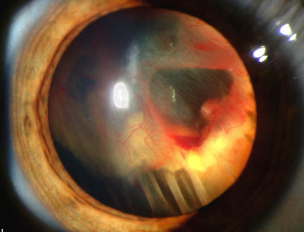
The retina is a very delicate organ of the eye. It is in charge of receiving light from the outside of the eye and transformation of light into electrical impulses which are then send via the optic nerve to the optic center in the brain. These electrical impulses are there perceived as light and actually processed and eventually recognized as certain objects.
The retina is not bigger than a post stamp. It has a central area (the macula) which is essential for central vision. There are two types of cells in the retina, cones and rods. While cones are responsible for sharpness of vision as well as color vision, rods are necessary for seeing in conditions of reduced illumination. The organ may be affected by many conditions and one is retinal detachment. It is characterized by a separation of the retina from the underlying tissues it is connected to i.e. is lying on.
Cataract Surgery and Retinal Detachment
Cataract surgery may initiate retinal detachment. The risk of retinal detachment increases even more if there are other complications after the surgery. However, since today many ophthalmologists opt for extracapsular surgery in patients suffering from cataract, the risk of retinal detachment is not so high.
The previous approach known as intracapsular cataract surgery included removal of the entire lens. The posterior part of the capsule of the lens was removed as well. This part is of great importance to keep the vitreous gel in its place and prevent retinal detachment. So, by adopting extracapsular approach when removing the cataract, surgeons leave the posterior part of the capsule of the lens intact and it does not allow vitreous detachment and subsequent retinal detachment.
Retinal Detachment Surgery and Potential Complications
If retinal detachment has occurred, the person must be treated surgically. Namely, the goal of the surgery is to move the retina to the back of the eye and fix it there, not allowing to detach again. This is achieved with an older method called scleral buckling and the newer one called pneumatic retinopexy.
Potential complications associated with retinal detachment surgeries include transient discomfort, watering, redness, itching and swelling and more complex problems like double vision, glaucoma, bleeding into the vitreous, retina and behind the retina. Furthermore, there may be drooping of the eyelid, infections and sometimes scleral buckle may need to be removed.Retinal Detachment Surgery Results
The surgery is successful in approximately 80% of all patients and if the procedure is repeated, the success reaches 90%. It may take up to several months until vision is completely restored. In patients who have experience detachment of the macula, central vision will never be the same as prior to detachment. Still, this procedure is considered highly efficient when it comes to restoring vision that would be otherwise completely lost.


Your thoughts on this
Loading...