Neck and Head Lymph Nodes
The role of every lymph node is to filter the lymph from various parts of the body. Our body contains around 500 to 600 lymph nodes localized all over the body.
Cervical or neck lymph nodes are divided into specific groups. The majority of cervical lymph nodes are available for palpation, and even the patient may notice if they become enlarged.
The rough division of lymph nodes can be into anterior and posterior groups of cervical lymph nodes. The anterior cervical lymph nodes are placed deep inside at the back of the neck, and they are in charge of drainage of the throat structures, thyroid gland, and tonsils. The posterior cervical lymph nodes are localized at the back of the neck along the front edge of the largest muscles in the neck. They can be palpated by the doctor. Their enlargement is easily noticed even by the patient.
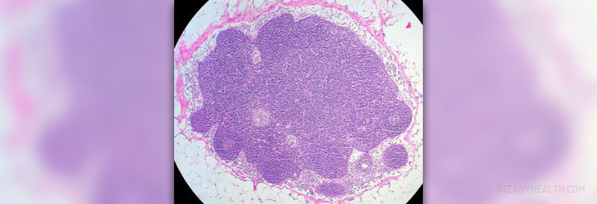
Submandibular lymph nodes are located below the angles of the mandibular bone, while a submental lymph node is located below the middle of the mandibular bone. They drain the lymph from the floor of the mouth.
Tonsilar lymph nodes drain tonsils and the back part of the pharynx.
Supraclavicular lymph nodes are located above the collar bone, and they can get enlarged in the majority of medical conditions, both benign and malignant.
Painful Lymph Nodes of the Neck
If lymph nodes of the neck become enlarged and painful, it is definitive that something is going on in the human body. Infections are in the majority of cases responsible for enlargement of cervical lymph nodes. Viral infections such as Epstein Bar virus infection can cause huge enlargement of cervical lymph nodes. Cervical lymph nodes may be additionally enlarged in bacterial infection, such as streptococcal angina. Even neglected problems with teeth, including cavities, may eventually result in enlargement of the neck lymph nodes.
Apart from infections, the reaction of the cervical lymph nodes may be connected to certain tumors. Namely, Hodgkin and non-Hodgkin lymphomas regularly lead to enlargement of lymph nodes, and cervical lymph nodes are affected in most cases. Cervical lymph nodes may be also enlarged due to the secondary spread of the cancer in the oral cavity, throat, thyroid gland, or larynx. Even in the case of stomach or lung cancer, metastases may affect cervical lymph nodes.
- A normal sized lymph node is usually less than one cm in diameter. Of course, there are exceptions in lymph nodes in different regions and at different ages have different sizes. For example, some authors have proposed that an inguinal lymph node size up to 1.5 cm should be considered normal, while the normal range for the epitrochlear nodes is up to 0.5cm.
- In general, normal lymph nodes are larger in children (ages 2-10), in whom a size of more than 2 cm is suggestive of a malignancy (i.e., lymphoma) or a granulomatous disease (such as tuberculosis or cat scratch disease).
- Ultrasound is a noninvasive method to assess lymph nodes in superficial regions like the neck. Computed tomography (CT) is useful to determine LAP in the thorax or abdominopelvic cavity. Tissue diagnosis by fine needle aspiration biopsy or excisional biopsy is the gold standard evaluation for LAP.
- Seventy-five percent of all LAPs are localized, with more than 50% being seen in the head and neck area. LAP may be localized or generalized.
Viral and bacterial infections are easily treated. However, if the treatment does not lead to a reduction in the size of the lymph nodes the doctor may suspect in malignant disease. The patient is further examined. The assessment includes laboratory tests, chest x-ray, CT scan of the head and neck, and finally, an MRI of the head and neck. The doctor will perform the biopsy of the enlarged lymph node, and the final diagnosis can be set after the pathophysiological evaluation of the biopted lymph node.








-Causes,-Symptoms,-Diagnosis,-Treatment_f_280x120.jpg)


Your thoughts on this
Loading...