Inguinal Lymph Nodes - Overview
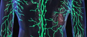
Lymph is a clear fluid that circulates inside the lymphatic system. The lymph flows through the lymph nodes, where it is filtrated from all the waste products. Lymph nodes are omnipresent and can be found in each part of the body. They drain the lymph from the specific body parts, and according to the body part they are classified into several groups. Inguinal lymph nodes are only one group of lymph nodes.
The inguinal lymph nodes can be classified into the superficial and deep inguinal nodes. The superficial inguinal lymph nodes are located below the inguinal ligament and deep to Camper's fascia. This fascia overlies the femoral vessels.
In humans, there are approximately 10 lymph nodes. These lymph nodes collect the lymph from the integument of the penis, scrotum, perineum, buttock, abdominal wall (below the level of umbilicus), vulva, anus, and lower extremity, and drain it to the deep inguinal nodes. The deep inguinal lymph nodes lie under the cribriform fascia, a bit medial to the femoral vein. In humans, there are 3 to 5 deep inguinal nodes.

Enlargement of Inguinal Lymph Nodes
Enlargement or swelling of the inguinal lymph nodes must be taken seriously since they normally cannot be palpated, and swelling of any kind only points to the presence of some underlying medical condition. There are numerous reasons why the inguinal lymph nodes enlarge. Once the patient notices the enlargement, he/ she is due to report it to the doctor.
The doctor will perform a physical examination and certain tests and identify the underlying cause. After a definitive diagnosis is set the doctor will choose and suggest the most suitable treatment. In some cases, enlargement of the inguinal lymph nodes is found accidentally during examination for other purposes.
The leading cause of inguinal node swelling is infection. A variety of infections, such as skin infections of the leg and lower abdominal wall, sexually transmitted diseases, bubonic plague, and many more are only several causes of inguinal lymph node enlargement. These lymph nodes may be also enlarged in a variety of autoimmune diseases, including rheumatoid arthritis and systemic lupus erythematosus.
Finally, swelling of the inguinal lymph nodes may be related to cancer. Primary cancer of lymph nodes (lymphoma) or the spread of the cancer to other organs may lead to enlargement of the inguinal lymph nodes.
No matter what the cause is, it is supposed to be identified as soon as possible. This way, the specific treatment can start on time and prevent potential complications of the primary disease.
- Among patients undergoing follow-up after surgery for melanoma, ultrasound (US) very often reveals lymph nodes in groin area, that do not show clear characters of a metastatic lesion yet that have atypical US features, which could result in diagnostic uncertainty. We evaluated such lesions among a cohort of patients.
- The study population consisted of patients who presented consecutively to our facility for a control between 1 January 2009 and 30 July 2010 and who had undergone surgery for a melanoma, at least 6 months earlier, in areas draining to lymph nodes of the groin but choosing – for this study - the opposite side to the natural drainage.
- The following parameters of the US performed on the lymph nodes were evaluated: number and size, aspects of the outline, including any extroflexion of the outline and contours morphology, homogeneity and thickness of the cortex and aspects of the hilus, characteristics of the vascularisation of the lymph node at color-power Doppler. A second US examination was performed on the same area after at least 12 months.
- We found a very high number of patients (42/124) with lymph nodes that did not appear to be fully normal at US examination, particularly those with structural alterations in the hilus and slight loss of physiologic curvature of the outlines, with moderate thickening of the cortex. Of the 124 patients, who were followed for at least one year, 42 showed these characteristics, and none of these showed any progression to malignancy at follow-up.


Your thoughts on this
Loading...