Primary congenital glaucoma is a condition present at birth and is most commonly diagnosed at the same time or a bit after the birth. In some cases the diagnosis can be set during the first year of life or even later in infancy and early childhood. The basic characteristic of primary congenital glaucoma is an inadequate development of the eye's drainage system. Inappropriate drainage of the aqueous fluid eventually leads to increased intraocular pressure and accompanying symptoms and signs of the disease.
In around 75% of all cases primary congenital glaucoma is bilateral. The condition predominantly affects boys. It is essential to diagnose the disease on time since only this way serious visual impairment can be successfully prevented.
Causes of Primary Congenital Glaucoma
The actual cause of primary congenital glaucoma is inadequate development of the eye's drainage channels. Since eye fluid simply cannot be drained properly it accumulates inside the eye and eventually causes increase in intraocular pressure. This increase of intraocular pressure can be tolerated up to certain extent but if it lingers for certain period of time it may cause serious damage to the optic nerve.
Presentation of Primary Congenital Glaucoma
This medical condition features with three characteristic signs. They include hypersensitivity to light (medically known as photophobia), excessive production of tears and spasms of the eyelids. These symptoms are easily noticed and once they have occurred the parents are supposed to take their child to a doctor.
Diagnosing Primary Congenital Glaucoma
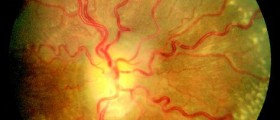
Primary congenital glaucoma is easily diagnosed by a well experienced ophthalmologist. The child undergoes special eye exams and tests including tonometry, examination and measurement of the cornea, gonioscopy, ophthalmoscopy and fundus photograph. All the previously mentioned together with typical signs of the disease help in setting of the definitive diagnosis.
Treatment for Primary Congenital Glaucoma
Primary congenital glaucoma is treated surgically. Medications are sometimes prescribed but they can only temporary lower increased intraocular pressure. The surgery solves the problem for good by making specific structural changes in the draining system of the affected eye. So the goal of the surgery is to allow the excess of intraocular fluid to be properly drained.
Surgeons most common perform goniotomy and trabeculotomy. In goniotomy the surgeon cuts certain structures and eliminates the resistance to fluid flow. Trabeculotomy requires a probe which creates perforation at the specific site this way allowing the excess of fluid to be drained. Both of the procedures are approximately successful. Only if these procedures fail, a surgeon performs additional procedures such as drainage implant surgery or ciliary body destructive procedures.









Your thoughts on this
Loading...