Losing a breast due to breast cancer is very traumatic for many women. Even though, some women are suitable to lumpectomy others have to face mastectomy and the loss of entire breast. For many years surgeons have tried to develop suitable reconstructive techniques which will increase women's self esteem and help them with their loss. DIEP flap microsurgery is one of the procedures which help in reconstruction of the lost breast.
DIEP Flap Microsurgical Breast Reconstruction
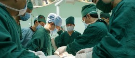
A DIEP flap is a type of surgery in which the skin, fat and accompanying blood vessels are transplanted from the abdomen to the chest wall. To be more precise, this transplantation is actually the first stage in total breast reconstruction. The goal of the surgery is to preserve muscles and motor nerves as well as all the vital structures in the transplant. Blood vessels of the flap are reconnected to the blood vessels in the chest wall or those located in the axilla. Reconnection of the blood vessels is performed under a microscope. Depending of the previous condition one or both breasts can be reconstructed this way. The entire procedure lasts approximately 7 hours and is performed under general anesthesia.
In stage 2 of the reconstruction the breast undergoes aesthetic shaping. In stage 2 the reconstruction of the nipple is completed. Stage 2 may also include any of the necessary counterbalancing procedures of the remaining breast such as breast reduction, augmentation or breast lift. This surgery is generally performed 3 months after the first surgery and it lasts no more than a couple of hours.
The terminal part of the reconstruction is areola reconstruction and is performed after two months.
Complications and Failure of DIEP Flap Microsurgical Reconstruction
Normal reaction to the surgery includes mild bilging of the abdominal wall. The risk of abdominal wall weakness and hernia is minimal. The most common complications are seromas which are usually treated with needle aspiration.
Failure of the procedure is reported in less than 1% of cases. If it occurs the failure is associated with inappropriate flap blood vessel anatomy. It may also occur if the flap is deprived from adequate blood supply from the blood vessels in the chest and axilla. And finally, injury to the essential blood vessels of the flap may be a significant contributor to failure. Failure is diagnosed before the patient is discharged and it is treated with additional microsurgical flap procedure right after the first one or the patient undergoes another surgery 3 months after the initial one.


Your thoughts on this
Loading...