
Granuloma Annulare - Clinical Characteristics
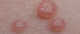
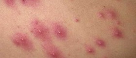
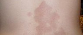
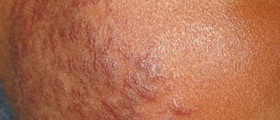
Skin lesions that affect people suffering from granuloma annulare develop in a form of nonscaling, dome-shaped or slightly flattened papule of approximately 3-6 mm in diameter. The skin changes are skin colored, pink or violaceous. The skin lesions on the lower extremities tend to be darker comparing to skin changes on the rest of the body. As the name of the condition suggests multiple papules are arranged in a form of a ring. These 'rings' are from 1 to 8 cm in diameter. Papules that form the border may not be fully confluent and they give 'beaded' appearance to the border. Each patient usually suffers from multiple rings. There is also a chance of fusion of the rings and this way a single larger lesion occurs. It is of a polycyclic configuration. Predilection places for granuloma annulare are the dorsal surface of the feet and the dorsal surface of the hands and fingers. Furthermore, extensor parts of the arms and legs such as the elbows and knees are commonly affected too. The condition may affect people of all ages but it is most frequent among children of age 4-10. The condition is easily confirmed with biopsy of skin lesions.
Unlike the typical clinical presentation of granuloma annulare the condition may develop in a form of so called atypical granuloma annulare. Namely, in this case the condition develops in a disseminated pattern and the skin lesions consist of hundreds of small rings. The entire surface of the body can be affected even though the rings predominantly affect sun-exposed surfaces. In children there is a chance for subcutaneous lesions that resemble rheumatoid nodules. In rather rare cases the skin changes undergo ulceration. This is a so called perforating granuloma annulare. And finally, in some cases the condition may simulate the appearance of necrobiosis lipoidica diabeticorum which makes differentiation of these two conditions very hard.
Course and Prognosis of Granuloma Annulare
The ring structures in the skin may change their shape over a period of weeks or months. In majority of cases the disease is self-limited and trace of the lesions usually disappear within 1-2 years. The actual cause of granuloma annulare is still unknown. The lesions resemble those that typically form in patients suffering from rheumatoid arthritis.
Treatment for Granuloma Annulare
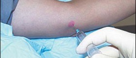
There is no effective treatment for granuloma annulare. Still, skin lesions may respond to certain medications such as high-potency topical steroids, particularly if they are used together with occlusion. Intralesionally injected steroids are more potent comparing to topical forms. The treatment is limited since it carries a risk of postinjectional atrophy.








Your thoughts on this
Loading...