Breast calcifications are small calcium areas located inside the breast. These are not palpable and can only be detected through X-ray or mammogram scanning. Fortunately, these growths are benign in most cases.
Types of Breast Calcifications
One type of breast calcifications are macrocalcifications, appearing like long dots or dashes on the scanned image. These are considered to be completely natural and common, affecting women as they age, once they are older than 50. Also, younger women can also have this form of breast calcifications, in some rare cases. The condition stems from calcium deposits in an existing cysts or milk ducts. Yet, injuries and inflammation of the area may result in the same phenomena. Macrocalcifications are considered to be harmless and, thereby, they do not require any special monitoring or interventions. Also, they cannot evolve into cancer.
Microcalcifications, manifesting through small white specks on the mammogram, usually in the areas where cells are replaced more quickly than what is considered to be common, are the other type of breast calcifications. These too are not considered malign in the vast majority of cases. Nevertheless, if many microcalcifications are seen in a single area, this could be a premonition of a breast cancer, being a malignant sign.
The Test
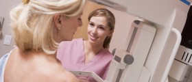
Since breast calcifications can be seen on a mammogram, this test is necessary for their diagnosis. A radiologist, who performs the scanning in the first place, will analyze the calcifications. Then, depending on the type, size, shape and other qualities of these growths, he/she will suggest further tests and exams. If macrocalcifications are the case, no treatment is necessary. However, in case of microcalcifications, you may need to have a close-up mammogram done. After this, your doctor may confirm that there is no cancer in the area, recommending no action to be taken or send you to a biopsy if the area is suspicious.
The biopsy involves extracting a piece of the calcified material in order to analyze it. This is commonly done with a needle, while the patient is under local anesthesia. Alternatively, a surgeon may remove larger area of the tissue which is then pathohistologically analyzed.
The test may show absence or signs of cancer. However, keep in mind that calcifications are benign in most cases, having no connections with cancer whatsoever. Nevertheless, if you are worried about this problem, have yourself tested for preventive purposes. Contact your nearest health care facility and seek further information on the subject.


_f_280x120.jpg)


Your thoughts on this
Loading...