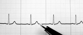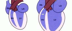
Right bundle branch block refers to the delaying or ceasing of electrical impulse transmission along the right bundle branch. This leads to the right depolarization of the right ventricle. This depolarization spreads from the interventricular septum and left ventricle to the opposing ventricle. Even though transmission along the right bundle branch might be interrupted, transmission in the left bundle branch might proceed normally. Depolarization of the left side ventricle and the left bundle branch occurs before the depolarization of the other side, which occurs relatively slowly.
Anatomy
The conduction system of the heart is made up of cells that transmit electrical impulses faster than the myocardium that surrounds them. The conduction system is divided into segments beginning at the AV junction and terminating with the Purkinje fibers. The right bundle branch originates at the septal leaflet of the tricuspid valve. It surfaces on the right ventricular septum just below the papillary muscle of the conus. At the terminal both this bundle branch (as well as the left bundle branch), Purkinje fibers are interlaced with the endocardial surface of the ventricle.
Causes, treatment and outlook
The most widely seen cause of RBBB with regard to children is surgical complication as a result of an isolated VSD repair. In up to 81% of this type of surgical operation, RBBB will occur. The onset of RBBB will be more likely in the case that the VSD is located close to the His-Purkinje system.
For the most part, this condition does not result in clinically problematic difficulties. Generally, the condition will follow a benign course, but in some cases it is possible for the condition to degenerate and lead to complete heart block and thus sudden cardiac death. This is often the case if the condition is accompanied by substantial injury to the His-Purkinje system.
Depending on the type of surgical repair that is undergone, some patients might be at risk of developing pulmonic valve insufficiency. Further to this, some patients might also suffer from residual stenosis of the pulmonary circulation. This will lead to significant exposure of the right ventricle to abnormal hemodynamics.
In general, there will be low mortality rates no further complications with the surgically-induced form of the condition. However, those who have undergone tetralogy of Fallot repair should be aware of the higher risk of ventricular arrhythmia and sudden death. If Brugada syndrom, ARVC or Kearns-Sayre syndromes have also occurred, one might also be at a higher risk of death.


Your thoughts on this
Loading...