
Peptic ulcers are growths which are greater than 10mm, affecting about 10% of adults in Western countries. One third of these ulcers are defined as gastric ulcers. A very small percentage of these ulcers can be malignant, being cancerous. Regardless, it is very important to rule out this characteristic through tests and analysis. Therefore, medical diagnosis and treatment of gastric ulcers is crucial.
Characteristics of Benign Gastric Ulcers
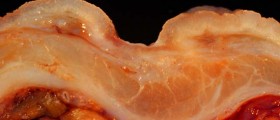
About 95% of all gastric ulcers are benign. A benign ulcer has some specific characteristics differentiating it from a malignant one. Firstly, it is projected beyond the healthy lumen, on the profile view. Secondly, the edges of the ulcer crater of this type are smooth and well defined, along with the filling that surrounds it.
If an ulcer seems to be lacking some or many of these characteristics, it needs to undergo further analysis through biopsy and endoscopy, since there is a chance that it is malignant.
Characteristics of Malign Gastric Ulcers
Contrary to the benign gastric ulcers, the malignant ones are located inside the lumen, except when an ulcer is located in the antrum or the greater curve. Next, the margins of these ulcers are not even and may contain nodules. Also, the mass around the ulcer is uneven and lacks symmetry. If an ulcer is found in the fundus, there are great chances that it is malignant.
Additional Facts Regarding Gastric Ulcers
When a gastric ulcer starts to heal, it actually decreases in size and becomes more linear than round. Usually, once a medical treatment commences, it takes about 8 weeks for the ulcer to completely disappear, provided that it is a benign one. When antral ulcers heal, this process may resemble a malignant one, often confusing doctors. If a lesser-curve ulcer starts healing, it can cause hourglass stomach retracting the abnormality of the opposite wall.
As far as the precision of scanning devices is concerned, single-contrast views are correct in 75% of gastric cancer diagnosis procedures. On the other hand, a double-contrast view is right in 95% of cases. Once an ulcer is perforated, CT scanning can prove to be an efficient method of monitoring the progress. Computed topography scan can be useful for these purposes as well.
Multidetector row CT scanning is an excellent method of diagnosing and monitoring stomach ulcers, since it provides images in 3D, giving accurate information regarding the growth, allowing doctors clear and precise insight. Virtual endroscopic imaging is yet another tool which is very useful for detecting gastric cancers, even in their primary stages of development. Finally, ultrasonography rules out gastric ulcers and notices gallstones or pancreatitis while sonographics show the outcomes of a perforated ulcer.









Your thoughts on this
Loading...