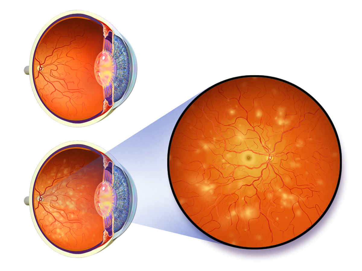
Diabetic retinopathy is the result of two processes regarding the blood vessels of the retina - blood vessel closure and abnormal vessel permeability.
Retinal blood vessel closure
The capillaries are usually the first to suffer from vessel closure. They are very important for healthy retina because they supply the oxygen and carry away carbon dioxide and waste. As the oxygen supply decreases to certain parts of the retina, the adjacent capillaries dilate, causing microaneurysms. These microaneurysms are visible as tiny red dots in an ophthalmologic exam.
Nonperfusion or localized closure of retinal capillaries is not easy to notice by the patient, unless it is in the center of the retina, where it reduces the vision. In more severe cases, retinal capillary closure can lead to proliferative diabetic retinopathy, in which new blood vessels are formed. These new vessels are fragile and bleed easily, leaving small scars on the retina.
In addition to capillary closure, small arteries or arterioles in the retina can also close off, leading to reduced blood supply and small, white dots or patches in the retina.
Abnormal vessel permeability
The blood vessels in the retina are different from those in other parts of the body. While most blood vessels have tiny openings that allows fluid to pass through the wall, retinal blood vessels have tight junctions between the cells in the wall, so the liquid has to pass trough the cells. In diabetic retinopathy, the blood vessel walls become more permeable, meaning that fluids, fats, proteins, blood cells and other larger molecules can pass through the walls and leak into the retina. If the fluid accumulates in the center of the retina, it is called macular edema or diabetic maculopathy. This leads to the formation of yellow clumps or spots called exudates. While the water gets absorbed quickly in the retinal tissue, fat takes a lot more time to be absorbed and leaves marks in form of rings.
In non-proliferative diabetic retinopathy, this swelling or edema is considered serious only if it is located at the areas where it causes difficulties with vision. If there is a need for it, it can be solved with laser eye surgery.
- www.cdc.gov/visionhealth/vehss/data/studies/diabetic-retinopathy.html
- www.cdc.gov/nhsn/pdfs/validation/2017/icd-10-cm-ddc.xlsx
- Photo courtesy of BruceBlaus by Wikimedia Commons: commons.wikimedia.org/wiki/File:Blausen_0312_DiabeticRetinopathy.png









Your thoughts on this
Loading...