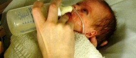
Encephalitis caused by the Herpes simplex virus (HSV) is very serious disease, which may end up with a significant brain damage or even death. This condition, more specifically HSV type 2, may affect newborn babies causing neonatal herpes and generalized infection of the brain. The other type of this disease is known to affect children over 3 months and adult people. In this case, HSV type 1 affects only temporal and frontal lobes of the brain.
HSV remains dormant in the nervous system (the neurons) and in rare occasions it gets transported through peripheral neurons to the brain (central nervous system), provoking encephalitis. HSV encephalitis is considered to be medical emergency, as this is the most serious complication of the HSV infection that exists.

How HSV Affects the Brain?
Doctors still don’t know everything about this virus and the way it causes encephalitis. However, neurons get destroyed very quickly in this disease, especially those located in the medical temporal and inferior frontal lobes. In rare cases HSV encephalitis can affect some other parts of the brain, such as cerebellum, basal ganglia or the brainstem.
Brain cells get damaged directly because of the virus and also because of the immune system of the body, responding to infection. Viral infection spreads through the brain, from one side to the other, provoking necrosis of the cells and inflammation.
About 30% of all herpes encephalitis (HSE) is caused directly because of the primary HSV infection, while other cases are usually associated with some preexisting HSV infection and its reactivation.
What to Expect from Herpes Encephalitis Diagnosis?
Herpes encephalitis is potentially fatal neurological disease and for that reason it must be treated. In untreated patients, HSE is a progressive problem and usually ends lethally after a week or two in some 70% of cases. Those who survive untreated HSV encephalitis may expect serious neurologic problems.
- Among common central nervous system (CNS) viral infections, mortality in HSE is disproportionately high, taking into account a recent study that showed that HSV infections are responsible for only 11% of cases compared with 29% for varicella-zoster virus, another human herpesvirus that is not associated with such a high mortality.
- The neuropathogenesis of HSE has intrigued clinicians and scientists for many years, with two of the key questions relating to, firstly, the very low incidence of the condition in the presence of the widespread carriage of latent HSV in ganglionic tissues in healthy people and, secondly, the propensity of the disease process to localise to the frontotemporal region. About 90% of normal people are seropositive for HSV-1 indicating past exposure to the virus, a finding that is consistent with the presence of latent HSV-1 genomes in the trigeminal ganglia of 85–90% of people at unselected necropsy.
- Regarding the site specificity of HSE, the pathway of viral spread is probably more important than cell-type viral susceptibility. The unique anatomical localisation has been thought to result from entry of the virus via the olfactory pathway with spread along the base of the brain to the temporal lobes, a view that is supported by the immunocytochemical evidence of HSV antigens in the olfactory tract and cortex, as well as temporal lobes, hippocampus, amygdaloid nucleus, insula, and cingulate gyrus in patients dying from HSE.
- The index of suspicion of HSE should always be high for a patient presenting with the typical features of encephalitis such as fever, headache, confusion, and clouding of consciousness.
- The diagnosis of HSE is usually established from the combination of the clinical and investigative features. Magnetic resonance imaging (MRI) provides the most sensitive method of detecting early lesions and is the imaging of choice in HSE; if MRI is available it should be the first diagnostic step after clinical assessment.
- Examination of the cerebrospinal fluid (CSF) is of considerable diagnostic value in HSE and should always be performed after computed tomography or MRI. The exception, in our view, is where cranial imaging shows evidence of severe cerebral oedema and brain shift, in which case we prefer to delay lumbar puncture until the oedema is reduced with steroids or mannitol because of the risk of brain herniation. While about 5% of patients have normal CSF, the characteristic profile consists of a normal or raised pressure, a lymphocytic pleiocytosis (typically 10–200 cells/mm3), normal glucose, and increased protein (0.6 to 6 g/l). Red blood cells and xanthochromia may be present in the CSF in some patients but are of no diagnostic value in distinguishing HSE from other causes of encephalitis.
- Brain biopsy in HSE now has only a limited role. Before the advent of acyclovir, brain biopsy was regularly used to make a definitive diagnosis of HSE, as both its sensitivity and specificity are extremely high (95% and > 99%, respectively). However, the advent of HSV PCR and acyclovir has resulted in few neurologists advocating a brain biopsy to diagnose HSE.
Acyclovir and vidarabine are drugs recommended for the treatment of this condition. However, doctors usually prefer acyclovir because of the studies which revealed just 19% (or 6 to 11%, depending on the studies) mortality rate. These results are much better than in patients treated with vidarabine. After the treatment with acyclovir, some 38% of the patients had no or just mild neurological deficits, 9% had moderate and 53% experienced severe neurological problems.
Neonatal HSE is quite often fatal medical problem. About 6% of babies suffering from isolated HSE and some 31% of those with disseminated HSV infection in the brain have fatal consequences.
Prognosis is also found to be much better for non-comatose patients, especially in those younger than 30 years of age, while coma usually means bad prognosis.


Your thoughts on this
Loading...