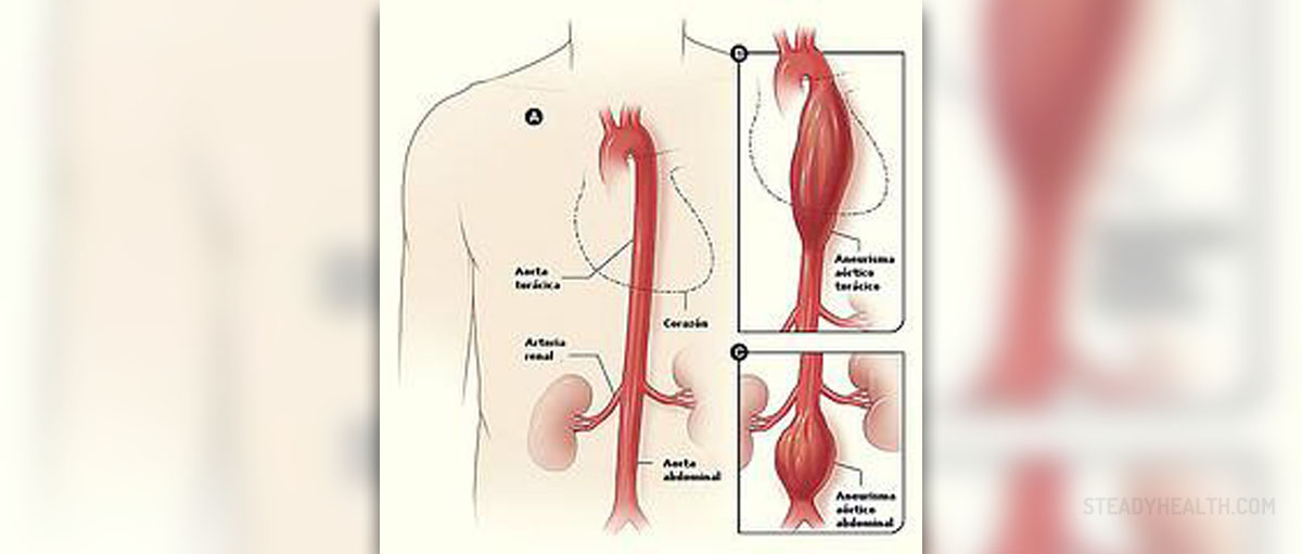
Aortic aneurysm is the medical term used for any bulging of an artery which distorts the appearance of the blood vessel and makes it susceptible to rupture. In order for an aneurysm to be classified as such it should dilate and reach at least 1.5 times size of the normal artery. The aorta is the leading blood vessels in the body affected by aneurysms. This particularly refers to its abdominal part while other portions such as the arch, the ascendens aorta or the thoracic aorta might be affected too but in rare instances. Brain arteries are also prone to aneurysms. All in all, the bulging is not benign, it carries the risk of rupture and also contributes to blood clot formation. This is why each and every aneurysm should be timely diagnosed, monitored and at some point surgically treated.
What is AAA and Who Gets It?
AAA is short for abdominal aortic aneurysm. This is actually a bulging of the abdominal part of the aorta, the one that runs from the lower surface of the diaphragm through the entire abdominal cavity down to the pelvis. There are many branches that separate from the blood vessels providing blood supply to various organs and tissues in the abdomen.
This localized ballooning of the aorta usually exceeds the normal diameter of the blood vessels by 50% or even more. Abdominal aortic aneurysm is practically the most reported type of all aneurysms. In the majority of cases (90%) the bulging develops bellow the kidneys i.e. occurs infrarenally. Pararenal and suprarenal location of abdominal aortic aneurysm is also reported. The aneurysm rarely stays the same size. In fact it tends to enlarge and the larger it is, the greater are the chances it will rupture.
Medical experts have estimated that individuals between 65 and 75 years or age represent the leading group of patients suffering from abdominal aortic aneurysm. Men seem to be affected more. The condition is also reported more among smokers. The aneurysm per se is generally asymptomatic. It may sometimes precipitate abdominal discomfort or pain in the back due to compression of nearby tissues and organs. What is more, pain might affect the legs as a result of disturbed blood flow.
Most surgeons agree that aneurysms larger than 5.5. cm in diameter must be operated. This is because the risk increases significantly once the bulging reaches this size. The rupture is the most severe complication blamed for many lethal outcomes. Namely, mortality even if patients reach the hospital is between 60 and 90%. Now, the surgery itself carries the risk of death. Such risk usually do not to exceed 1-6% which is much lower compared to the risk of rupture.
All in all, most experts agree on the necessity of screening of males over 65 years of age for abdominal aortic aneurysm. The exam is not complex, is noninvasive and might identify most of the aneurysms on time, preventing death in many individuals.
Ultrasound for Diagnosis
Patients who are suspected of suffering from abdominal aortic aneurysm first undergo a physical exam. A health care provider palpates the abdomen and while doing so he/she may easily see the pulsation of the abdominal wall while deeper palpation reveals the bulge inside the abdomen. It may be challenging to exam obese individuals in whom the fat in the abdominal area makes deep palpation practically impossible. So only extremely thin people have obvious pulsation while in obese individuals other diagnostic tool should be indicated. For instance, doctors use stethoscopes and auscultate the abdomen. The auscultation reveals a bruit of abnormal sound caused by turbulence of blood within the affected blood vessel.
Plain abdominal X- ray is one more approach to diagnosing abdominal aortic aneurysms. Namely, it can easily reveal calcium deposits in the aorta which are typical characteristic of the condition. However, X-ray is not precise and simply cannot provide with insight in the extent of bulging and the relation of the aneurysm and the nearby organs. For this purpose doctors use ultrasound.
Ultrasound has approximately 98% accuracy when it comes to measuring the size of the abdominal aortic aneurysm. This is the exam that gives a clear picture of the aorta, its structural changes, the size and shape of the aneurysm and potential compression of nearby tissues/organs. Ultrasound is noninvasive, safe and painless exam usually performed first. Still, surgeons require additional exams which will be even more precise when evaluating details regarding the bulging necessary for the surgery. This is achieved with CT scans of the abdomen. This imaging technique is highly sensitive and may provide with plenty of details regarding the aneurysm. Better visualization is achieved with intravenously administered contrast dye. The exam includes certain amount of radiation and patients allergic to contract dye should not receive this enhancing substance because of potential allergic reactions. Dyes are also forbidden in patients suffering from kidney disorders.
Sometimes, it is even possible to engage MRI for visualization of the abdominal aortic aneurysm. Both MRI and CT are suitable for diagnosing aneurysms but they last longer than ultrasound. On the other hand, these imaging techniques provide with more details of the changes affecting the aorta allowing surgeons to plan the surgery much better.






Your thoughts on this
Loading...