Transposition of the Great Vessels
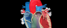
This medical condition includes transposition of two main arteries of the heart, the aorta and the pulmonary artery. The aorta is normally connected to the right ventricle and the pulmonary artery to the left ventricle. The function of the aorta is to carry the blood rich in oxygen while the pulmonary artery delivered the blood rich in carbon dioxide to the lungs where this blood is re-oxygenated. In transposition of great arteries the result is inappropriate oxygenation of the blood. The blood which is supposed to flow separately at some point mix and this leads to the symptoms and signs of the disease. This disease is congenital. This means that babies are born with it and the defect has occurred during intrauterine growth and development. The actual cause has not been found yet. Still, the only way for a baby to survive and to overcome the problem is surgical repair of the deformity.
Symptoms of Transposition of the Great Vessels
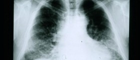
The problem of inappropriate oxygenation of the blood and mixture of the blood rich in oxygen and that rich in carbon dioxide result in cyanosis. The baby develops central type of cyanosis and its skin is entirely blue. The cyanosis is most prominent of the fingertips and the mouth. After the birth the doctor may auscultate heart murmurs and the chest and heart X ray examination will confirm a cardiac enlargement as well as pulmonary over circulation. Additionally, an oxygen monitor will report low levels of oxygen in the blood. And finally, suitable insight in the transposition can be achieved by echocardiography. The deformity may allow the baby to live for some time but only the surgery will save it from unavoidable lethal outcome.
Treatment for Transposition of the Great Vessels
The mortality rate of these children is rather high. Some of them will die within a month and only small number will live up to a year if not operated. In certain cases a surgical procedure called septostomy may help. It is performed by using cardiac catheterization. In some patients prostaglandin may be given in order to open or expand the small tube between the aorta and the pulmonary artery.
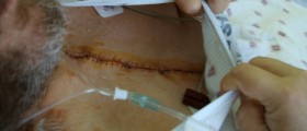
There are two surgical approaches which are used. In the first one a surgeon makes a tunnel in the upper chambers in order to redirect the blood rich in oxygen and the blood rich in carbon dioxide into the correct direction. This procedure is performed in rather young infants.
Another option is detachment of the major arteries from the heart and the correct replacement of each of them. The aorta and the pulmonary artery are eventually attached to the correct ventricle. The best results are achieved if this procedure is done a few days or weeks after the birth.
The results of both procedures are excellent and the baby continues to live normally.







Your thoughts on this
Loading...