
Inherited disorders
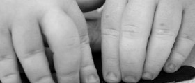
All of these disorders are present at birth, spread out and not considered to be progressive. At this moment, the researchers have discovered almost 200 distinct disorders that can all be described. Out of all these disorders, two are seen more often than any other disorder. These disorders are named X-linked recessive hypohidrotic ectodermal dysplasia and hidrotic ectodermal dysplasia.One of the main characteristics of ectodermal dysplasias are certain abnormalities. These abnormalities can occur in various ectodermal structures. Not all of these disorders are of the same magnitude as some can be meek while others can be crushing. Diagnosing these conditions is not an easy task for the doctors as a lot of the abnormalities do not develop until the baby has become an infant or even a child. Dental, hair and nail abnormalities are the ones that are not apparent until that age is reached. Parents who suffer from any of the abnormalities should mention that to the doctor as that will ease the diagnosis. Any family member who is diagnosed with ectodermal dysplasias increases the chance of the infant being born with them. Apart from the obvious signs of EDs, there are other signs like hyperthermia accompanied by fever and seizures, conjunctivitis, hearing and vision that starts to decrease, dental carries that occurs more often than it is normal, mental retardation, dysphagia, improper growth and otitis, rhinitis and pharyngitis that occur quite a lot. These are not all the symptoms.
The experts are still unsure about all the causes of ectodermal dysplasias and the genetic defects are known in no more than 30 disorders. The good thing is that the cause of one of the EDs that occurs most often was discovered. Certain mutations in EDA are the cause of x-linked recessive hypohidrotic ectodermal dysplasia. These mutations decipher the ectodysplasin protein and that protein plays a vital role in development of the cells and their endurance and distinction. Mutations in other genes are the factors that lead to ectodermal dysplasias. For instance, mutations in the EDARADD gene lead to autosomal recessive hypohidrotic ectodermal dysplasia.Classification of ectodermal dysplasia’s
The experts have come up with the classification of ectodermal dysplasias. They have classified them according to the clinical characteristics. However, pure EDs and ectodermal dysplasias are not defined in the same way. While they are characterized by abnormalities in nowhere else but in the ectodermal structures, ectodermal dysplasias syndromes are classified by ectodermal defects that are connected with other anomalousness. The original classification of ectodermal dysplasias first appeared in 1982 and it was created by Freire-Maia and Pinheiro. In year 1994 and again in 2001 the classification was updated. The classification system placed ectodermal dysplasias in subgroups based on either the presence of the lack of hair anomalies, dental abnormalities, nail abnormalities and eccrine gland dysfunction.All of the ectodermal dysplasias were classified into either A group disorders or B group disorders. Group A consists of defects in no less than 2 original ectodermal construction, while group B is made of a defect in one of the structures along with a defect in some other ectodermal construction like the ears or lips. The experts have come up with eleven subgroups for group A disorders.
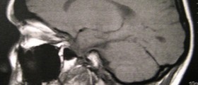
However, with the experts being able to identify the actual causes of ectodermal dysplasias, new classifications are being made. Ectodermal dysplasias were classified again in 2003, where the experts have placed the disorders into four groups according to the inherent pathophysiologic fault. The groups are cell-to-cell communication and signaling, adhesion, development and other. Before this one, another classification was made and it consisted of only two groups, defects in developmental regulation and defects in cytoskeleton maintenance and cell stability.Laboratory studies do not play an important role when it comes to diagnosing and treating these disorders. However, imaging studies do play a significant role in identifying the defects. Certain skeletal malformations that are present in hands or feet can be seen on an x-ray. Apart from an x-ray, orthopantography and renal ultrasound are useful as they can identify dental abnormalcies and discovering inherent genitourinary tract anomalousness, if there are any. In addition to these, other tests like sweat pore counts, pilocarpine iontophoresis and skin biopsy can be helpful as well. The number of eccrine glands is being looked at by these tests.




Your thoughts on this
Loading...