
Dermatofibroma is a benign skin tumor made of fibrous tissue. It originates from dermal dendritic histiocyte cells. The tumor may form at the site of a minor skin injury (e.g. after an insect bite). Scientists have not managed to identify the exact cause of why this tumor occurs.
Dermatofibroma Clinical Characteristics
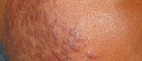
The tumor predominantly develops on the legs and arms. Once dermatofibroma is formed, it generally stays forever since it has no tendency to withdraw spontaneously. This benign skin lesion is in a form of firm-feeling nodule, yellow-brown in color although there have been cases of pink and even dark colored dermatofibromas. Dark dermatofibromas are often found in dark colored individuals. If the skin lesion is squeezed, the skin above the tumor becomes dimpled.
In many cases there is only one tumor. However, some individuals may develop more than one tumor and there have even been cases when dozens of dermatofibromas have erupted within a few months. Multiple eruption is basically associated with immunosuppression (patients suffering from cancer, people treated with corticosteroids or other immunosupressive medications).
Dermatofibroma is usually asymptomatic and causes no additional symptoms apart from being visible. Still, some people may complain about pruritus (persistent itch of the affected skin) and tenderness of the area.
How Frequent is Dermatofibroma?
These skin lesions are considered relatively common in the Unites States. The same data are collected in other countries as well. The occurrence of tumor among different races is similar, but the tumor still predominantly affects women. It can affect people of all age.
Even though dermatofibroma is generally classified as benign tumor, there have been the few case reports of the occurrence of metastatic dermatofibroma. Still, this is rather challenging diagnosis as well as disputable from the standpoint of pathohistologic diagnosis. In the reported cases tumors have been highly cellular, larger and they have shown local recurrence.
Dermatofibroma Diagnosis and Treatment
In order to confirm or rule out diagnosis of dermatofibroma doctors perform biopsy of the tumor (a skin biopsy). There are several pathohistological types of dermatofibroma (cellular, aneurismal, epitheloid, atypical, lipidized ankle-type, palisading and cholesterotic). It is essential to differentiate the tumor from a skin cancer, especially dermatofibrosarcoma and desmoplastic form of malign melanoma.
Since dermatofibroma does not cause any discomfort it may not be treated at all. It is more an aesthetic problem. There have been attempts to remove the tumor with liquid nitrogen, intralesional steroid injections and surgical procedures such as inverted pyramidal biopsy, superficial shaving and cryosurgery. However, none of them is very successful. Laser treatment is used in people suffering from multiple dermatofibromas, particularly those located on the face.






Your thoughts on this
Loading...